Published - Thu, 04 Aug 2022

HOW TO EVALUATE ILL PEDIATRIC PATIENT

HOW TO EVALUATE ILL PEDIATRIC PATIENT

1. A primary survey should be performed immediately.

a) Airway: The patency of the airway needs to be evaluated.

— To remove tongue obstruction, the airway should be freed with external techniques.

— Obstruction from foreign bodies should be recognized and relieved.

— Gas exchange should be assessed and optimized.

— Aspiration of gastric or oral contents should be prevented.

— An artificial airway should be provided if no protective reflexes exist.

b) Breathing: The adequacy of ventilation should be assessed, including the equality of chest rise and breath sounds.

— A determination of whether air entry is impaired from a central or pulmonary cause should be made.

— Ventilation should be enhanced if necessary with mouth-to-mouth breathing, bag-mask ventilation, or endotracheal intubation with or without bag assistance.

— A surgical airway should be provided if an oral or endotracheal airway cannot be established.

c) Circulation should be assessed, including the quality and intensity of pulses in the upper and lower extremity and blood pressure.

— Hemorrhages should be controlled with direct pressure until surgical intervention can be accomplished.

— Intravenous vascular access and a resuscitative fluid bolus should be provided; if intravenous access cannot be rapidly achieved, intraosseous access should be performed.

— External cardiac massage should be provided if no pulse exists.

— Pharmacologic management of circulation should be provided if the fluid status is adequate and cardiac output is impaired because of either vascular or pump instability.

— Conversion of dangerous cardiac rhythm disturbances should be provided if hypotension and unresponsiveness ensue.

d) Neurologic examination: A preliminary assessment should be made according to the following simple scale:

— Alert and responsive to verbal stimuli

— Responsive to verbal stimuli but no spontaneous alertness.

— Responsive only to painful stimuli.

— Unresponsive to any stimuli

e) Overall examination:

— Infants should be unclothed and examined rapidly for injuries.

— Passive heat loss should be prevented by removing wet clothing and by providing external warmth.

— The patient’s core temperature should be obtained to monitor for hypo- or hyperthermia.


2. A secondary survey, entailing a detailed physical examination and history, is then performed.

a) Head

— Evidence of trauma is determined by palpating bony prominences and the maxillae and by inspecting the nose and ears for drainage of cerebrospinal fluid (CSF).

— The existence of dehydration is determined by inspecting the mucosae for evidence of decreased tearing or moisture or sunken eyes.

— The eyes should be inspected for pupillary size and extraocular movements. A funduscopic examination should be performed if possible to assess for the central nervous system (CNS) or toxic involvement.

— The oral cavity should be inspected for odor or discoloration that may imply a toxicologic basis for the condition.

b) Neck

— The neck should be palpated for midline tenderness or deformity.

— Flexion, extension, and rotation should be performed only after an injury have been excluded either clinically or by radiographic means.

c) Chest

— The chest wall should be inspected for symmetric expansion or disarticulation.

— Palpation for change in respiratory fremitus or inequality should be performed.

— Auscultation of the chest for breath sounds and evidence of adventitial sounds such as rales, rhonchi, or wheezes should be performed.

— Penetrating or blunt chest trauma is treated immediately.

d) Abdomen

— The abdominal wall should be inspected for bruising, penetration, or hematoma.

— Palpation for trauma, masses, or guarding and rigidity, which may indicate peritonitis, should be performed.

— Palpation of the flanks to search for hematoma or an expanding mass should be performed.

e) Pelvis

— The pelvis should be palpated laterally and in an anteroposterior position for tenderness or instability.

— The perineum should be inspected for lacerations, active bleeding, or hematoma. The urethra and rectum should be inspected for trauma or blood.

— In age-appropriate females, a pelvic and rectal examination should be performed, and pregnancy should be excluded.

f) Extremities

— The extremities should be inspected for signs of abrasion, hematoma, laceration, or deformity.

— Palpation of the bones for instability, tenderness, or deformity should be performed.

g) Neurologic assessment

— Attention should be focused on cranial nerve function, motor function, and sensory function, along with the patient’s level of consciousness.

— The presence or absence of reflexes should be noted.

— Serial examinations for changes in the level of consciousness or loss of neurologic function should be performed.


3. Additional studies are often nonspecific but may provide clues to the diagnosis.

a) Laboratory studies

— Aerobic and anaerobic cultures of blood

— Arterial blood gas (ABG) determination

— Complete blood count (CBC) and differential

— Electrolytes and bicarbonate

— Blood urea nitrogen and creatinine

— Immediate fingerstick glucose and serum glucose

— Urinalysis and urine culture

— Stool culture

b) Radiologic studies

— Abdominal radiography

— Abdominal ultrasonography

— Chest radiography

— Computed tomography (CT) of the head/abdomen

c) Ancillary studies

— Echocardiography

— Electrocardiography

— Lumbar puncture

— Urine for toxicologic analysis


Comments (0)

Search
Popular categories
Latest blogs
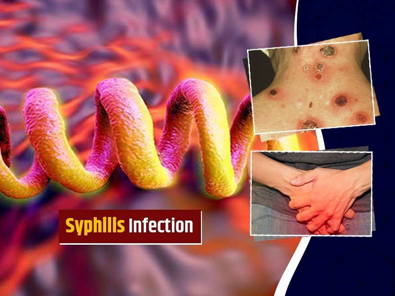
All you need to know about Syphilis
ETIOLOGY: Syphilis is caused by T. pallidum.INCIDENCE: Each year, there are 29,000 new cases of syphilis. This figure represents probably only 10% of actual cases.CLINICAL FEATURES1. Primary syphilis: After an average incubation period of approximately 3 weeks, a smooth, painless ulcer called a chancre appears at the site of primary inoculation. The chancre heals without treatment in approximately 3 to 6 weeks; at about the same time, a painless uni- or bilateral regional adenopathy develops.2. Secondary syphilis represents disseminated disease and occurs in all patients with untreated primary infection. The lesions of secondary syphilis are papulosquamous lesions that occur over the entire trunk, extremities, penis, and buttocks. Fever and weight loss occur in 70% of patients.3. Tertiary syphilis occurs at least 10 years after the primary infection in at least 30% to 35% of untreated patients. The two most important manifestations of tertiary syphilis are cardiovascular syphilis, causing thoracic aneurysms, and neurosyphilis, causing meningitis, stroke, seizures, dementia, general weakness, and posterior column dysfunction.DIFFERENTIAL DIAGNOSES— Chancroid— HSV type 1 infection— Lymphogranuloma venereum— Tinea, sarcoid— Lichen planus— Seborrhea dermatitis— Molluscum contagiosum— Traumatic ulcer— Furuncle— CarcinomaEVALUATION: The clinical diagnosis can be confirmed by darkfield microscopic examination or more commonly serologic testing.THERAPY1. The standard treatment for primary, secondary, and early tertiary syphilis is benzathine penicillin G (2.4 million U administered intramuscularly as a single dose).2. For late tertiary syphilis or neurosyphilis, benzathine penicillin G (2.4 million U, three doses administered intramuscularly 1 week apart) is used. Doxycycline (100 mg orally twice daily for 14 days) can be given to patients who are allergic to penicillin.DISPOSITION1. Primary and secondary syphilis can be treated on an outpatient basis.2. Patients with neurosyphilis or major cardiovascular manifestations require admission for intravenous therapy.

Tue, 15 Nov 2022

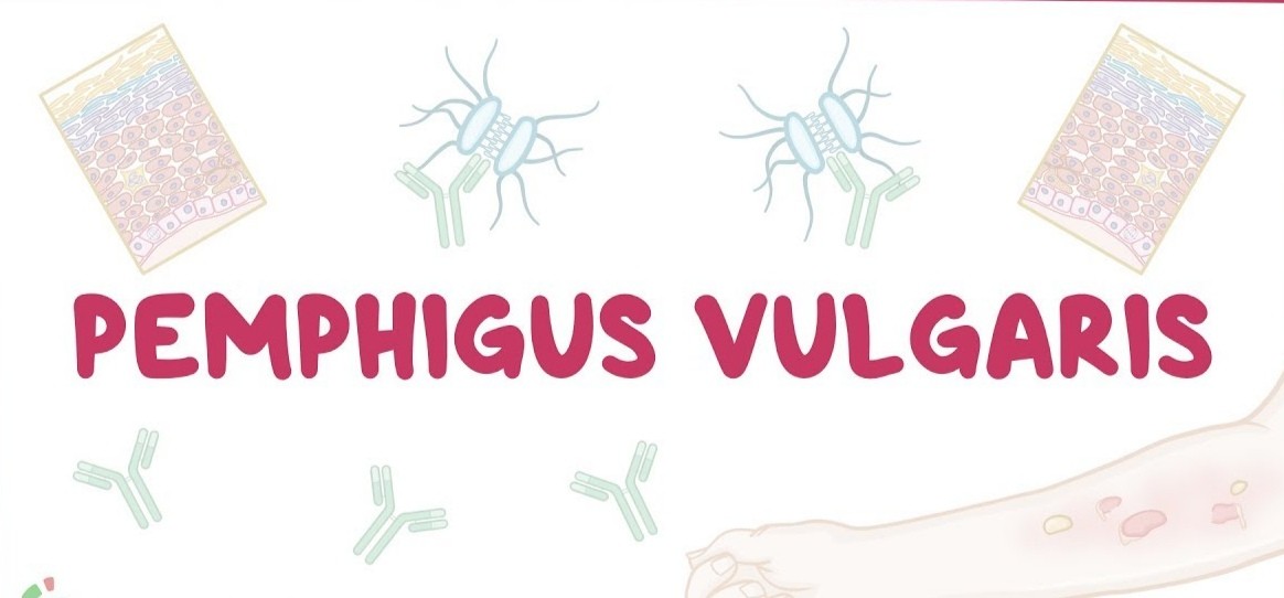
What is Pemphigus Vulgaris?
Pemphigus Vulgaris is a rare disease that affects elderly patients. The mortality rate is 10%; most deaths result from steroid complications, secondary infection, dehydration, or thromboembolism. Pemphigus Vulgaris is caused by the attachment of immunoglobulin G autoantibodies to the epidermis. It has been associated with D -penicillamine and captopril administration.CLINICAL FEATURES1. Mucosal lesions and erosions are very common. Examination of all mucosal sites is warranted.2. Non-pruritic, painful, flaccid bullae appear that rupture easily. Blisters can be extended or new bullae formed by applying firm tangential pressure on the intact epidermis.3. Weakness, weight loss, and dysphagia may be presenting complaints.DIFFERENTIAL DIAGNOSES— Erythema multiforme— Bullous impetigo— Herpes zosterEVALUATION: Biopsy of lesions shows eosinophils, intraepidermal bullae, and acantholysis. Indirect immunofluorescent staining shows immunoglobulin G antibodies. Serum titers can be followed to evaluate the effectiveness of therapy.THERAPY1. Prednisone (200 to 350 mg/day) for 5 to 10 weeks is used until the cessation of new blister formation occurs. The dosage is then reduced to 40 mg on alternative days and tapered over 1 year.2. Azathioprine (100 mg/day) is added to the regimen and the dosage is reduced over a 4- to 6-month period. Methotrexate and cyclophosphamide can be used instead of azathioprine.3. Topical analgesics (e.g., viscous lidocaine) can be used to alleviate the pain associated with oral lesions.DISPOSITION: Patients with severe cases and oral lesions may require hospital admission for intravenous hydration. Others can be treated as outpatients with close follow-up.

Tue, 15 Nov 2022

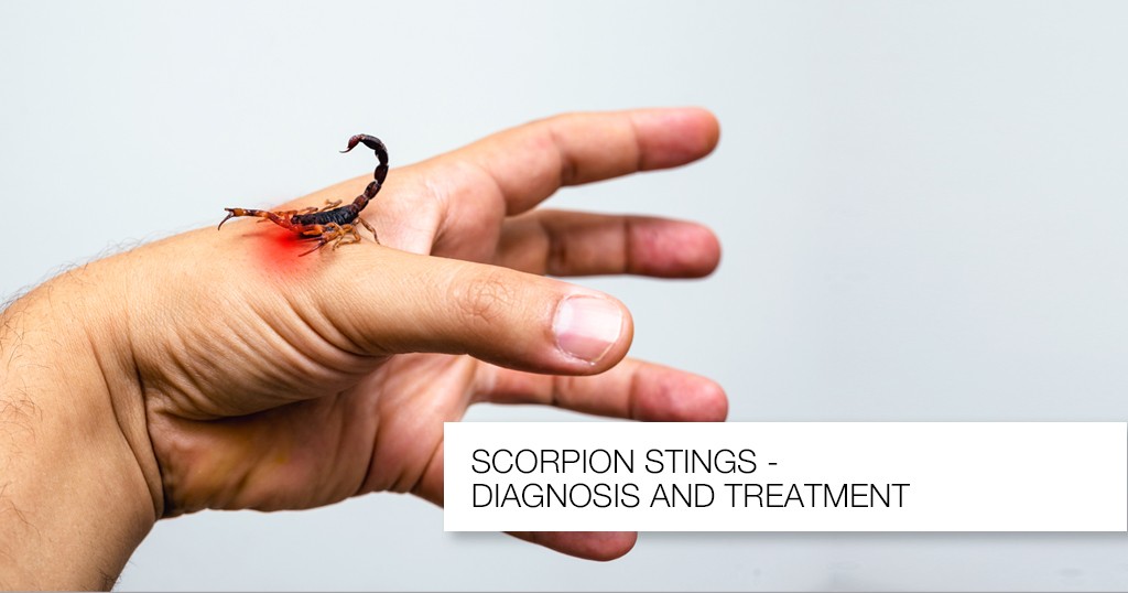
Know about Scorpion Stings
Introduction: A nocturnal arachnid that lives in the Southwest of the United States is the scorpion. It has two venom glands and a stinger in its tail. The majority of species are rather benign, and they often only create a limited reaction similar to that brought on by a bee sting. The bark scorpion (Centruroides sculpturatus) venom, on the other hand, has neurotoxic that can result in a serious reaction. This dangerous scorpion is found on trees in Arizona and New Mexico.CLINICAL FEATURES1. Local effects: The C. sculpturatus scorpion bite causes immediate, excruciating pain at the stung site, as well as swelling and ultimately, numbness. The area that was hurt is extremely sensitive, and the implicated extremity could become paralyzed.2. Systemic effects: The neurotoxin is strongly cholinergic and can cause excessive salivation, blurred vision, muscular spasms, hypertension, and respiratory difficulties.DIFFERENTIAL DIAGNOSES — Snakebite— A puncture wound or other trauma— Insect or spider bite— Drug intoxicationEVALUATION: Typically, the offending scorpion is seen or assumed by history; if safety allows the scorpion to be brought in, this is best. Due to the wide range of symptoms and quick progression, a thorough history and physical examination are necessary.THERAPY1. Pre-hospital management includes rapid transportation of the patient, application of an ice pack to the sting site, and safe transport of the scorpion for identification. When serious symptoms appear, life-saving procedures should be started.2. ED managementa) Antivenin should be administered in all cases of severe envenomation.b) Ventilatory support may be required, with intubation and oxygen for patients with severe systemic response or anaphylaxis.c) Atropine may be required to counteract the cholinergic effects; the dose is titrated to relieve the cholinergic signs.d) Benzodiazepines may be used for seizures and muscle spasms.DISPOSITION: All victims should be observed for 24 hours, especially children. Symptomatic patients should be transferred to the intensive care unit if symptoms are severe.

Sat, 12 Nov 2022

All blogs