Created by - Dr. Prashant Raj
Management and Treatment of Pleural Effusion
Transudative effusions are managed by treating the underlying medical disorder. However, despite whether it’s a transudative or exudative, large, refractory pleural effusions inflicting severe symptoms need to be drained to produce symptomatic relief. Treatment for PLEURAL effusion focuses on removing the additional fluid from the space and preventing it from rebuilding again. The goal is to alleviate symptoms and treat any underlying medical conditions that are causing the fluid build-up. One of the foremost common procedures to get rid of additional fluid is termed thoracentesis. This involves exploration by ultrasound to find the fluid and a hollow needle is inserted to empty the fluid from the space. Thoracentesis will improve respiratory, scale back coughing and improve oxygen levels. It is very common for fluid to build up again after it is removed, thus patients typically would need more than one procedure. If that is the case, your doctor could counsel a patient into another procedure known as pleurodesis where mild inflammation is deliberately created between the lung and chest cavity after removing the excess fluid by injecting a drug which would cause the sticking of layers of the pleura.Medications Medications could also be accustomed treat serosa effusion betting on its cause and symptoms. forms of medications used could include: Antibiotics if there's an infection Steroids and anti-inflammatory drugs to alleviate pain and scale back inflammation or swelling Diuretics assist the body to eliminate additional fluid through urine. Bronchodilators widen the airways within the lungs and permit air to flow easily Surgery Occasionally, surgery could also be needed to treat pleural effusion, particularly if it continues to come back. Pleurodesis may be a procedure that seals the layers of the pleura Pleurectomy may be a procedure that removes a part of the pleural membrane to forestall continuing fluid buildup.
More detailsPublished - Wed, 26 Oct 2022

Created by - Dr. Prashant Raj
How to Approach the Patient with Dermatologic Lesions
PATIENT HISTORY: Answers to the following questions should be sought:1. General questions: Duration, changes over time, exacerbating/alleviating factors, travel history?2. Questions about symptoms: Itching, painful, swelling, change in color, associated symptoms?3. Questions about the past medical history: New medications, occupational and social history, family history?PHYSICAL EXAMINATION: The patient’s entire body should be examined, and the lesion should be palpated.1. Location of lesionsa) Site: The location of the lesions on the body may offer a clue to the diagnosis. For example, varicella spares the palms and soles, and scabies favors the finger web spaces.b) Extent: Are the lesions localized or generalized?c) Arrangement: Are the lesions grouped or solitary?2. Appearance of lesionsa) Margins can be raised (as in psoriasis) or more active peripherally (as in tinea).b) Color: Melanin and hemoglobin are responsible for the colors of lesions.c) Shape: Annular lesions have clear or contrasting centers (e.g., the target lesions of Stevens-Johnson syndrome).d) Scale: The epidermis is replaced every 28 days. In psoriasis, the basal cells are mitotically active and produce an epidermis or scale.TYPES OF LESIONSa) Macules are flat lesions, measuring less than 1cm in diameter that are different in color from the normal skin tone (e.g., measles).b) Papules are circumscribed, palpable elevations of the skin measuring less than 1cm in diameter (e.g., scabies).c) Nodules are circumscribed, palpable elevations of the skin measuring greater than 1 cm in diameter (e.g., erythema nodosum).d) Patches are flat lesions greater than 1cm in diameter (e.g., pityriasis rosea).e) Plaques are raised lesions measuring greater than 1cm in diameter (e.g., psoriasis).f) Pustules are raised lesions greater than 0.5 cm in diameter that contains yellow fluid (e.g., abscesses).g) Vesicles are raised lesions up to 0.5 cm in diameter that contains clear fluid (e.g., herpes simplex).h) Bullae are vesicles that are greater than 0.5 cm in diameter (e.g., pemphigus).i) The crust is a dried exudate (e.g., impetigo).DIFFERENTIAL DIAGNOSES: Many systemic diseases have cutaneous manifestations. For example:1. Necrobiosis lipoidica diabeticorum, an oval, yellowish, shiny plaque with sharp borders, is a characteristic lesion usually located on the shins of patients with diabetes.2. Erythema nodosum lesions may be seen in patients with ulcerative colitis.3. Erythema multiforme can be associated with viral infections such as herpes simplex.EVALUATION1. Microscopic examination: An ordinary microscope can be used to examine scrapings from skin lesions.a) A potassium hydroxide (KOH) slide preparation is used when dermatophyte infections are suspected. The slide is examined for long, thin, branching hyphae.b) A Tzanck slide preparation can confirm the diagnosis of herpes infections. Vesicle contents are smeared on a slide, and Wright’s stain or methylene blue is used for staining. Alternatively, the slide can be sent for direct fluorescent assay or enzyme-linked immunosorbent assay.2. Wood’s light examination: A Wood’s light is an ultraviolet lamp that emits radiation at a wavelength of 360 nm.a) In the presence of certain species of tinea capitis infection, the hair shaft will fluoresce bright green.b) Cutaneous corynebacterial infections fluoresce coral pink.3. Skin biopsy can be performed with basic surgical tools (e.g., forceps, scalpel, curette, scissors, and a needle holder) and is best performed by a dermatologist. A specific section of the lesion must be biopsied to obtain a satisfactory specimen for pathology. In addition, the specimen may need to be sent for immunofluorescence studies, electron microscopic examination, or culture.
More detailsPublished - Thu, 27 Oct 2022

Created by - Dr. Prashant Raj
Important indication for Diabetes
It is necessary to grasp that there are 2 major sorts of polygenic disorder and that they every have totally different causes. regardless of the cause, the main drawback in polygenic disorder is that the body is unable to utilize glucose. The result's prolonged elevations of blood glucose within the blood, that eventually wreaks disturbance on several organs, just like the eyes, kidney, heart, brain, and blood offer to the extremities.CausesFamily history -Family history is a vital risk issue for sort two polygenic disorder. If either one among the oldsters has the disorder, then one or all the kids can also develop sort two polygenic disorder later in life. This risk isn't one hundred pc secure and may be reduced by creating way changes. Ethnicities -Certain ethnicities are a lot of liable to developing polygenic disorder than others. for instance, the best risk of polygenic disorder is in African Americans, and therefore the lowest risk is in Caucasians. However, different ethnicities at high risk for polygenic disorder embrace the following:• Asians• Native Americans• Hispanic Americans• Pacific islanders Inherited DisorderSome genetic disorders, like bronzed diabetes and CF, will harm the exocrine gland and increase the danger of polygenic disorder. as luck would have it, each CF and bronzed diabetes isn't that common. Age -Advancing age could be a risk issue for sort two polygenic disorder. With age, all the organs within the body can impede and become inefficient. a particular share of seniors, particularly those that are weighty and lead an inactive life, are terribly seemingly to develop sort two polygenic disorder. Unhealthy Diet- Perhaps one among the most important risk factors for sort two polygenic disorder is associate unhealthy diet. several people these days consume quick foods and processed foods often. These foods ar high in calories, contain saturated fats, and have high meat content, that ar all risk factors for weight gain, resulting in polygenic disorder. the most effective recommendation is to eat a plant-based diet that has fruits, veggies, whole wheat, low-fat dairy farm, and nuts.
More detailsPublished - Thu, 27 Oct 2022

Created by - Dr. Prashant Raj
Treatment method of Type 1 Diabetes
Type 1 Diabetes polygenic disease could be a womb-to-tomb condition. The body doesn't manufacture enough hypoglycaemic agent, whereas blood glucose levels stay high unless an individual uses medication to manage them. The symptoms of kind one polygenic disease sometimes seem. They include:• increased hunger and thirst• frequent voiding• blurred vision• tiredness and fatigue• weight loss while not a plain trigger or cause Children, the primary signs are those of diabetic acidosis (DKA). this can be a probably serious condition wherever too several ketones flow into within the body, resulting in pathology. It wants immediate medical attention.Symptoms include:• a fruity smell on the breath• dry or flushed skin• nausea or reflex• abdominal pain• breathing problem• confusion and problem focusingManaging kind one polygenic disease will facilitate scale back the chance of assorted complications.These include:• diabetic retinopathy, which might cause vision loss Trusted supply• diabetic pathology, that affects nerve perform• problems with wound healing, particularly as nerve issues will create it tougher to note wounds• diabetic uropathy (kidney disease), which might cause kidney disease• cardiovascular sickness, coronary failure, stroke, and peripheral tube sickness• dental issues, as well as tooth loss andgum sickness• mental health issues, like depression The main treatment Trusted supply for kind one polygenic disease is hypoglycemic agent. individuals will take it using:• a needle and syringe• an hypoglycemic agent pen• an hypoglycemic agent pumpIf hypoglycemic agent doesn't absolutely management aldohexose levels, some individuals may have extra mediation, like Pramlintide (Symlin), that helps manage aldohexose levels when feeding. A doctor can advise on the simplest choice for every person.
More detailsPublished - Fri, 28 Oct 2022

Created by - Dr. Prashant Raj
How Can You Help Someone With Anxiety And Panic Attacks
Anxiety disorder is characterized by feelings of uneasiness, danger, tension, stress, irritability, or fatigue. Panic disorder is a form of anxiety that is characterized by recurrent panic attacks (i.e., sudden episodes of intense fear or impending doom associated with a variety of somatic symptoms).1. Panic attacks are often unpredictable, although they may occur commonly in certain situations.2. First-time panic attacks are rarely seen in patients older than 35 years; organic disorders should be considered in these cases.CLINICAL FEATURES: Symptoms associated with both generalized anxiety and panic attacks may mimic life-threatening conditions but include palpitations or pounding heart, diaphoresis, sensation of shortness of breath or choking, chest pain or discomfort, nausea, dizziness, light-headedness, numbness, tingling, or chills.DIFFERENTIAL DIAGNOSES: Many serious medical conditions can mimic the symptoms of panic disorder and need to be considered (Acute myocardial infarction, Cardiac arrhythmias, Pulmonary emboli, Hyperthyroidism and thyroid storm, Pheochromocytoma, Mitral valve prolapse, Alcohol withdrawal Use of central nervous system stimulants, Hypoglycemia, Asthma exacerbation, Stroke or serious intracranial abnormalities), especially in patients older than 45 years.Evaluation1. Patient history: A careful history of recurrent, short-lived episodes of panic will usually lead to the correct diagnosis of panic attacks. Anxiety may be harder to determine but is often a diagnosis of exclusion in the ED.2. Laboratory studies should be guided by the patient history and physical exam as well as age and presentation and may include a serum electrolyte panel and glucose level, and a drug screen. Older patients or those with atypical symptoms should have a thorough workup to detect conditions on a medical basis.3. Electrocardiography and radiology: An electrocardiogram or chest radiograph may be warranted.Therapy: Generally, both medications and behavioral therapy are used for patients with anxiety and panic disorders. Usually, medical therapy for panic attacks should be started by the patient’s primary care physician or a psychiatrist, not in the ED. However, commonly used medications include the following:1. Selective serotonin reuptake inhibitors are first-line therapy for the long-term control of anxiety and panic attacks.2. Benzodiazepines are both useful for panic attacks and usually produce a response within several hours to days of beginning the medication but should be used sparingly due to abuse and withdrawal potential.3. Disposition: Most will be discharged, although patients with risk factors for the significant disease should be admitted to the hospital to rule out a life-threatening condition.
More detailsPublished - Fri, 28 Oct 2022

Created by - Dr. Prashant Raj
Unusual Causes of Hyperthermia
1. Malignant hyperthermia: When under general anaesthesia, some patients with a rare genetic tendency may quickly experience acute hyperthermia, stiffness of the muscles, and acidosis. Inappropriate intracellular calcium release results in malignant hyperthermia. Dantrolene, which reduces myoplasmic calcium, is used as part of the treatment along with cooling and supportive measures.2. Neuroleptic malignant syndrome: This rare syndrome is induced by antipsychotic medications (e.g., haloperidol) and manifests as muscular rigidity, severe dyskinesia, dystonia, hyperthermia, dyspnea, tachycardia, and urinary incontinence. The brain's dopamine receptors are blocked as part of the technique. Additionally suppressing thirst, haloperidol makes the issue worse. Treatment includes supportive and cooling measures, with possible medications including dantrolene, amantadine, bromocriptine, and benzodiazepines.3. Drug overdose: Hyperpyrexia that is lethal can be brought on by anticholinergic medication overdose and sympathomimetic drugs like amphetamines.4. Cerebrovascular accident: Ischemic or hemorrhagic strokes involving the thermoregulatory centers in the brain can cause elevation of body temperature and should be considered in the differential diagnosis of hyperthermia.
More detailsPublished - Sun, 30 Oct 2022

Created by - Dr. Prashant Raj
Aortic Stenosis: Causes, Symptoms & Treatment
CAUSE: Aortic valve stenosis arises from defective valvular architecture. The ordinary dynamic stress of blood flow across the defective valve progressively traumatizes the valve, resulting in thickening, calcification, and narrowing of the valve orifice.1. Congenital: A congenital bicuspid valve is the cause of aortic stenosis in 50% of symptomatic patients.2. Rheumatic endocarditis leads to commissural fusion of valve leaflets, often affecting the mitral valve as well.3. Degenerative calcific aortic stenosis occurs in elderly patients and appears to be part of the aging process. Degenerative calcific aortic stenosis is less likely to result in symptoms.PATHOPHYSIOLOGY1. The obstruction to ventricular outflow that results from the stenotic valve stimulates concentric hypertrophy of the left ventricle to overcome the systolic pressure gradient and maintain cardiac output. 2. The increased muscle mass of the ventricle leads to increased myocardial oxygen demands. The hypertrophied hyperdynamic ventricle loses its ability to compensate for hemodynamic changes and eventually fails, leading to increased atrial pressures and pulmonary congestion. 3. The ventricle also loses its ability to increase cardiac output, leading to syncope or angina with exertion. CLINICAL FEATURES1. Typically, symptoms don't appear until the valve orifice has shrunk to less than 1cm2. Patients may have been diagnosed previously or may present for the first time to the ED with dyspnea, angina, or syncope.2. Physical examination findings: The carotid arterial pulse takes longer and has less energy. The maximum impulse point could be hyperdynamic and enlarged. Auscultation of the heart reveals a harsh systolic murmur that occurs just after the S1 and is transmitted to the carotid arteries. The S2 may diminish as the disease progresses and the contribution of the aortic component (A2) is lost.DIFFERENTIAL DIAGNOSES The murmur of aortic stenosis must be differentiated from other systolic murmurs such as occur with mitral regurgitation, tricuspid regurgitation, pulmonic stenosis, and hypertrophic cardiomyopathy. Significant aortic stenosis must be differentiated from insignificant flow murmurs.EVALUATION1. Electrocardiography: Most patients show electrocardiographic evidence of left ventricular hypertrophy.2. Radiography: Before the development of severe aortic stenosis, a chest radiograph is typically normal. Later on, this could show indications of CHF and an enlarged cardiac silhouette.THERAPY: The presenting complaint directs the management of emergencies. Many of these individuals will eventually require valve replacement or repair.DISPOSITION: Patients with syncope, cardiac chest pain, CHF, or arrhythmias usually require admission to the hospital.
More detailsPublished - Tue, 01 Nov 2022

Created by - Dr. Prashant Raj
Obstructive Urinary Retention
The inability to urinate causes urinary retention, which causes the bladder to swell. Both blockage and non-obstruction are potential causes of urinary retention. Your urinary tract becomes blocked and urine cannot flow freely if there is an impediment (such as bladder or kidney stones). This is the cause of acute urine retention, which could be fatal.CAUSES1. Prostate enlargement: Urinary retention in adult men is often due to an enlarged prostate gland.2. Urethral strictures 3. Urethral foreign bodies 4. Paraphimosis and phimosis CLINICAL FEATURES1. Symptoms: Urinary symptoms include hesitancy, frequency, nocturia, urgency, and a decrease in the amount and force of the urine stream, resulting in “dribbling.”2. Physical examination findings include increased pain to suprapubic palpation and dullness to percussion over the distended fluid-filled bladder.DIFFERENTIAL DIAGNOSES — Neurogenic causes— Drug-induced retention— Urinary retention secondary to pain— Psychogenic causes— Renal failure— Abdominal aortic aneurysm— Bowel obstruction— A gravid uterusEVALUATION1. Physical examination readily identifies an obstructive etiology. A rectal examination should be performed to identify prostatic enlargement.2. Urinary catheterization: A postvoid residual of more than 300 mL of urine confirms the diagnosis.3. Laboratory studies— Urinalysis should be performed to rule out infection.— Renal and electrolyte panel. Blood urea nitrogen, creatinine, and electrolyte levels should be assessed to rule out renal insufficiency.4. Imaging: Ultrasound can reveal a distended bladder. CT of the abdomen and pelvis is often obtained in search of a kidney stone or other potential cause (malignancy).THERAPY1. Acute relief: Catheterization should provide acute relief.2. Definitive therapy: The source of the obstruction should be treated.DISPOSITIONMost patients with mechanical obstruction may be discharged home with an indwelling catheter and a leg bag (and antibiotics). A follow-up with a urologist should be arranged.
More detailsPublished - Wed, 02 Nov 2022

Created by - Dr. Prashant Raj
Neurologic and Pharmacologic Urinary Retention
A. NEUROLOGICCAUSES: Urinary retention may occur secondary to spinal cord trauma or compression, neuropathy (e.g., multiple sclerosis, diabetes mellitus, Guillain-Barré syndrome, tabes dorsalis), or viral infection.CLINICAL FEATURES1. Spinal cord injury or spinal shocka) Areflexia (atonic) bladder is seen with spinal pathology at or below the second lumbar vertebra.b) Reflex (hypertonic) bladder is seen with pathology above the second lumbar vertebra.2. Spinal cord compression: Back pain, gait disturbance, and hyperreflexia accompanying urinary retention may be indicative of spinal cord compression (e.g., by a tumor, herniated disk, or abscess).DIFFERENTIAL DIAGNOSES— Drug-induced retention— Urinary retention secondary to pain— Psychogenic causes— Renal failure— Abdominal aortic aneurysm— Bowel obstruction— A gravid uterusEVALUATION: A CT scan or magnetic resonance imaging scan should be obtained to evaluate spinal cord trauma or suspected compressive etiologies.THERAPY1. Acute reliefa) Atonic bladder may be relieved with self-catheterization or urecholine therapy.b) Hypertonic bladder may be relieved with self-catheterization or flavoxate or oxybutynin therapy.2. Definitive therapy depends on the underlying cause. Consultation with a specialist (e.g., a neurosurgeon, orthopedic surgeon, or neurologist) may be necessary.B. PHARMACOLOGICCAUSES:— Antihistamines— Antidepressants— α-adrenergic medications, including over-the-counter preparations, and many illicit drugs.DIFFERENTIAL DIAGNOSES— Urethral obstruction— Neurogenic retention— Urinary retention secondary to pain— Psychogenic causes— Renal failure— Abdominal aortic aneurysm— Bowel obstruction— A gravid uterusEVALUATION1. Patient history: A thorough patient history, including recently prescribed medications, often suggests the etiology.2. Physical examination should rule out other causes.THERAPY: Catheterization will provide acute relief. The offending medication should be discontinued following consultation with the patient’s primary care physician.
More detailsPublished - Fri, 04 Nov 2022
Search
Popular categories
Latest blogs

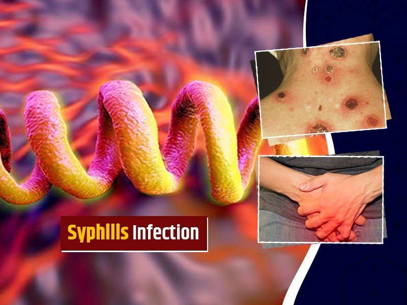
All you need to know about Syphilis
Tue, 15 Nov 2022

What is Pemphigus Vulgaris?
Tue, 15 Nov 2022

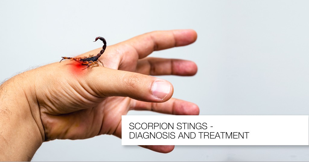
Know about Scorpion Stings
Sat, 12 Nov 2022
Write a public review