Latest blogs
Created by - Dr. Prashant Raj
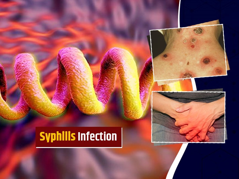
All you need to know about Syphilis
ETIOLOGY: Syphilis is caused by T. pallidum.INCIDENCE: Each year, there are 29,000 new cases of syphilis. This figure represents probably only 10% of actual cases.CLINICAL FEATURES1. Primary syphilis: After an average incubation period of approximately 3 weeks, a smooth, painless ulcer called a chancre appears at the site of primary inoculation. The chancre heals without treatment in approximately 3 to 6 weeks; at about the same time, a painless uni- or bilateral regional adenopathy develops.2. Secondary syphilis represents disseminated disease and occurs in all patients with untreated primary infection. The lesions of secondary syphilis are papulosquamous lesions that occur over the entire trunk, extremities, penis, and buttocks. Fever and weight loss occur in 70% of patients.3. Tertiary syphilis occurs at least 10 years after the primary infection in at least 30% to 35% of untreated patients. The two most important manifestations of tertiary syphilis are cardiovascular syphilis, causing thoracic aneurysms, and neurosyphilis, causing meningitis, stroke, seizures, dementia, general weakness, and posterior column dysfunction.DIFFERENTIAL DIAGNOSES— Chancroid— HSV type 1 infection— Lymphogranuloma venereum— Tinea, sarcoid— Lichen planus— Seborrhea dermatitis— Molluscum contagiosum— Traumatic ulcer— Furuncle— CarcinomaEVALUATION: The clinical diagnosis can be confirmed by darkfield microscopic examination or more commonly serologic testing.THERAPY1. The standard treatment for primary, secondary, and early tertiary syphilis is benzathine penicillin G (2.4 million U administered intramuscularly as a single dose).2. For late tertiary syphilis or neurosyphilis, benzathine penicillin G (2.4 million U, three doses administered intramuscularly 1 week apart) is used. Doxycycline (100 mg orally twice daily for 14 days) can be given to patients who are allergic to penicillin.DISPOSITION1. Primary and secondary syphilis can be treated on an outpatient basis.2. Patients with neurosyphilis or major cardiovascular manifestations require admission for intravenous therapy.
More detailsPublished - Tue, 15 Nov 2022

Created by - Dr. Prashant Raj
What is Pemphigus Vulgaris?
Pemphigus Vulgaris is a rare disease that affects elderly patients. The mortality rate is 10%; most deaths result from steroid complications, secondary infection, dehydration, or thromboembolism. Pemphigus Vulgaris is caused by the attachment of immunoglobulin G autoantibodies to the epidermis. It has been associated with D -penicillamine and captopril administration.CLINICAL FEATURES1. Mucosal lesions and erosions are very common. Examination of all mucosal sites is warranted.2. Non-pruritic, painful, flaccid bullae appear that rupture easily. Blisters can be extended or new bullae formed by applying firm tangential pressure on the intact epidermis.3. Weakness, weight loss, and dysphagia may be presenting complaints.DIFFERENTIAL DIAGNOSES— Erythema multiforme— Bullous impetigo— Herpes zosterEVALUATION: Biopsy of lesions shows eosinophils, intraepidermal bullae, and acantholysis. Indirect immunofluorescent staining shows immunoglobulin G antibodies. Serum titers can be followed to evaluate the effectiveness of therapy.THERAPY1. Prednisone (200 to 350 mg/day) for 5 to 10 weeks is used until the cessation of new blister formation occurs. The dosage is then reduced to 40 mg on alternative days and tapered over 1 year.2. Azathioprine (100 mg/day) is added to the regimen and the dosage is reduced over a 4- to 6-month period. Methotrexate and cyclophosphamide can be used instead of azathioprine.3. Topical analgesics (e.g., viscous lidocaine) can be used to alleviate the pain associated with oral lesions.DISPOSITION: Patients with severe cases and oral lesions may require hospital admission for intravenous hydration. Others can be treated as outpatients with close follow-up.
More detailsPublished - Tue, 15 Nov 2022
Created by - Dr. Prashant Raj
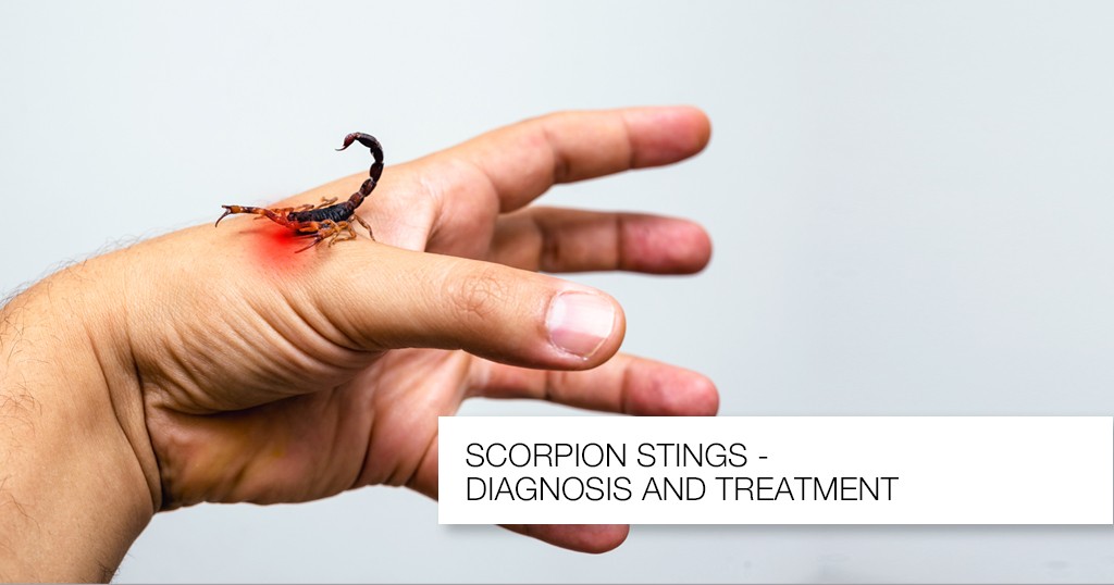
Know about Scorpion Stings
Introduction: A nocturnal arachnid that lives in the Southwest of the United States is the scorpion. It has two venom glands and a stinger in its tail. The majority of species are rather benign, and they often only create a limited reaction similar to that brought on by a bee sting. The bark scorpion (Centruroides sculpturatus) venom, on the other hand, has neurotoxic that can result in a serious reaction. This dangerous scorpion is found on trees in Arizona and New Mexico.CLINICAL FEATURES1. Local effects: The C. sculpturatus scorpion bite causes immediate, excruciating pain at the stung site, as well as swelling and ultimately, numbness. The area that was hurt is extremely sensitive, and the implicated extremity could become paralyzed.2. Systemic effects: The neurotoxin is strongly cholinergic and can cause excessive salivation, blurred vision, muscular spasms, hypertension, and respiratory difficulties.DIFFERENTIAL DIAGNOSES — Snakebite— A puncture wound or other trauma— Insect or spider bite— Drug intoxicationEVALUATION: Typically, the offending scorpion is seen or assumed by history; if safety allows the scorpion to be brought in, this is best. Due to the wide range of symptoms and quick progression, a thorough history and physical examination are necessary.THERAPY1. Pre-hospital management includes rapid transportation of the patient, application of an ice pack to the sting site, and safe transport of the scorpion for identification. When serious symptoms appear, life-saving procedures should be started.2. ED managementa) Antivenin should be administered in all cases of severe envenomation.b) Ventilatory support may be required, with intubation and oxygen for patients with severe systemic response or anaphylaxis.c) Atropine may be required to counteract the cholinergic effects; the dose is titrated to relieve the cholinergic signs.d) Benzodiazepines may be used for seizures and muscle spasms.DISPOSITION: All victims should be observed for 24 hours, especially children. Symptomatic patients should be transferred to the intensive care unit if symptoms are severe.
More detailsPublished - Sat, 12 Nov 2022
Created by - Dr. Prashant Raj
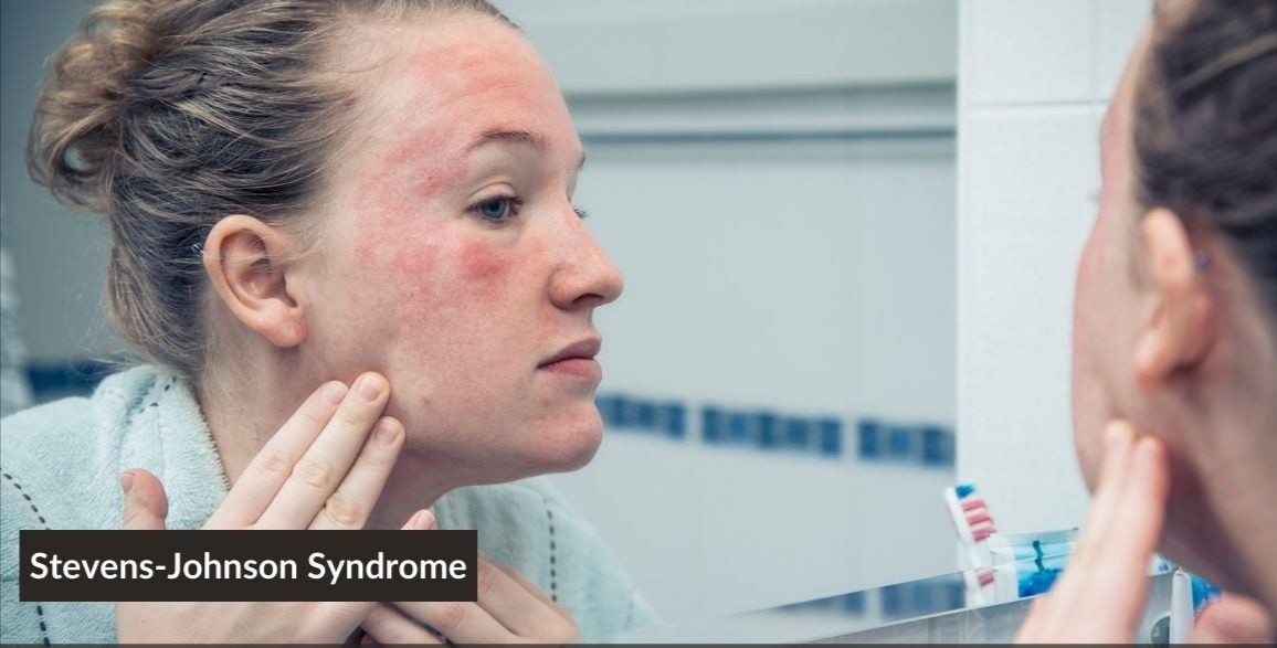
All you need to know about Stevens-Johnson Syndrome
Stevens-Johnson syndrome is a severe form of erythema multiforme, associated with a mortality rate of 10%. Complications include blindness, renal failure, meningitis, necrotizing tracheobronchitis, dehydration, secondary bacterial infection, arrhythmias, and congestive heart failure.Who is commonly affected?Children and young adults are most frequently affected by the disease. CLINICAL FEATURES1. Upper respiratory tract infection, headache, fever, hematuria, diarrhea, and arthralgias can precede the rash.2. Rash: Skin lesions are burning but not itchya) Bullae appear in 1 to 14 days on the skin and the mucous membranes of the mouth, genitalia, and anus.b) Ulcers: Corneal ulcers can lead to blindness. Ulcerative stomatitis leads to hemorrhagic crusting. Patients are unable to eat or drink and continuously drool.c) Vesicles rupture and leave denuded bases and necrotic epithelium.3. Signs of toxic epidermal necrolysis (TEN) may develop. Urinary retention can result from urethral involvement.DIFFERENTIAL DIAGNOSES include TEN and pemphigus.EVALUATION: Skin biopsy findings include necrolysis with edema and erythrocytes in the dermis.THERAPY1. Definitive treatment entails treating the cause if it can be identified (e.g., with antibiotics or termination of drug therapy). Prednisone (80 to 120 mg/day in divided doses) can be administered orally with subsequent tapering. Intravenous immunoglobulin has also been used successfully to halt the progression of TEN. It is important to note that treatment with prednisone or intravenous immunoglobulin is controversial and varies from one institution to another.2. Supportive treatmenta) Cool, wet compresses— Aluminum acetate compresses are applied to blisters.— Compresses soaked in potassium permanganate solution are applied to bullous lesions.b) Anesthetic troches, 2% viscous lidocaine, or 10% sodium bicarbonate mouthwashes can be used to soothe mouth lesions. If the patient cannot tolerate a liquid diet, he or she will require intravenous rehydration.c) Antibiotic therapy is indicated for patients with secondary bacterial infections.d) Ophthalmology consult: An ophthalmologist should be consulted.d) Urology consult: Urology should be consulted if genitourinary involvement is suspected.DISPOSITION: Patients with severe mucous membrane involvement require admission to a burn unit for reverse isolation and treatment of fluid and electrolyte imbalances.
More detailsPublished - Sat, 12 Nov 2022

Created by - Dr. Prashant Raj
Gonorrhea: Clinical Features, Evaluation & Treatment
ETIOLOGY: Gonorrhea is caused by Neisseria gonorrhoeae.Incidence: The ages of 20 and 24 have the highest frequency of the disease.CLINICAL FEATURES1. Local diseasea) Acute urethritis is the most common presentation in heterosexual men. Symptoms begin within 1 to 14 days of exposure and consist of dysuria and penile discharge. Three to ten percent of men with gonorrhea may be asymptomatic.b) Cervicitis: Primary gonorrhea in women is usually asymptomatic, and when symptoms do occur, they are usually mild and non-specific. Up to 20% of women with primary gonorrhea develop pelvic inflammatory disease, and 33% to 81% of women with the pelvic inflammatory disease have gonorrhea.c) Pharyngeal gonorrhea can be asymptomatic: The pharynx is colonized in 3% to 7% of heterosexual men, 5% to 20% of women, 10% to 25% of homosexual men, and 39% to 96% of pregnant women.d) Anorectal gonorrhea is common in both heterosexual women and homosexual men and is often asymptomatic. When symptoms occur, they are usually mild pruritus and rectal discomfort.2. Disseminated gonorrhea may complicate the disease course in 1% to 3% of patients with localized disease and is manifested most commonly as monoarticular arthritis or pustular dermatitis syndrome.DIFFERENTIAL DIAGNOSES1. Gonococcal urethritis must be differentiated from non-gonococcal urethritis caused by Chlamydia trachomatis.2. Disseminated gonorrhea: N. meningitidis infection, acute rheumatic fever, and Reiter syndrome must be ruled out. Differential diagnoses for skin lesions include syphilis, HIV infection, and condyloma acuminata.EVALUATION: Gram stain and culture of discharges are the cornerstones of diagnosis. All patients evaluated for gonorrhea should also have blood drawn for syphilis serology.THERAPY: For uncomplicated cervicitis or urethritis:1. Ceftriaxone (250 mg intramuscularly) and doxycycline (100 mg orally twice daily for 14 days, to cure possible concomitant Chlamydia infection) are the standard therapy.2. Cefixime (400 mg orally in a single dose) is no longer an accepted alternative due to resistance.3. Azithromycin, 2 g one-time dose, is an alternative for cephalosporin allergic patients.DISPOSITION1. Uncomplicated gonorrhea is managed on an outpatient basis. All sexual contacts must be identified and treated, and HIV testing should be considered by the patient with his or her primary physician at a later date.2. Patients with disseminated gonorrhea require hospitalization.
More detailsPublished - Fri, 11 Nov 2022

Created by - Dr. Prashant Raj
Alcoholic Ketoacidosis: Clinical Features, Evaluation & Treatment
Alcoholic ketoacidosis is usually seen in alcoholic patients who are forced to abruptly cease drinking alcohol after a drinking binge, but it may also be seen in first-time drinkers. Diabetes mellitus does not exist in these patients.PATHOGENESIS: The pathogenesis is uncertain. It is related to low insulin levels, reduction of available nicotinamide adenine dinucleotide, and increased ketone formation.CLINICAL FEATURES1. Patient history: The patient has recently stopped or limited alcohol consumption because of abdominal pain, nausea, and vomiting, not from a desire to stop drinking.2. Symptomsa) Diffuse abdominal pain is typically present. Pancreatitis, gastritis, and hepatitis, for example, are diseases linked to alcoholism that can induce abdominal discomfort. Other conditions unrelated to alcoholism might also produce abdominal pain (e.g., sepsis, pneumonia, pyelonephritis).b) There may be signs of alcohol withdrawal or delirium tremens.3. Physical examination findingsa) Hydration status: Dehydration occurs secondary to vomiting, diaphoresis, and decreased oral intake. The patient has tachycardia and hypotension and is critically unwell.b) Vital signs: Kussmaul respirations may be present, and the temperature may be elevated or normal.c) The range of mental states includes normal and comatose.d) Alcoholism stigmata, such as spider angiomata, may be seen.DIFFERENTIAL DIAGNOSES: An anion gap acidosis-causing condition needs to be ruled out. The most important conditions to take into account include isopropyl alcohol intoxication, hyperemesis gravidarum, hunger, cyanide poisoning, and diabetic ketoacidosis.EVALUATION1. Serum biochemical profile will establish the presence of an anion gap acidosis. A mixed disorder could also exist (e.g., metabolic ketoacidosis may occur from vomiting and respiratory alkalosis may occur from fever, sepsis, or alcohol withdrawal).2. Ketone studies: β-hydroxybutyric acid is the predominant ketone formed in alcoholic ketoacidosis. The nitroprusside test has limited use in individuals with alcoholic ketoacidosis since it only detects acetoacetate and not -hydroxybutyrate. Acetoacetate levels rise during treatment, giving the ketoacidosis a fictitious appearance of deteriorating.3. Bedside glucose determination: The level of blood sugar may be low, normal, or only slightly higher. Most individuals' blood glucose levels range from normal to elevated.THERAPY: Ketoacidosis can be reversed in 12 to 18 hours.1. Dehydration is treated with saline solutions containing glucose and thiamine. The clinical response appears to be enhanced by glucose. If inadequate oral intake is suspected, magnesium and vitamin supplements should be administered.2. Insulin: Administration of insulin is not indicated unless the patient has concomitant diabetes mellitus.3. Bicarbonate: Bicarbonate administration is under question. Most people recommend only giving bicarbonate in cases of cardiac arrest with known severe acidosis.DISPOSITION1. Admission: Patients with severe metabolic acidosis or those who are unable to tolerate oral fluids should be hospitalised. Prior to discharge, underlying or precipitating illnesses, as well as abdominal pain, must be assessed. Patients often react to therapy in 12 to 24 hours, at which point they may be released.2. Discharge: The patient might be released from the emergency room if the therapy goes well. It is crucial to closely monitor patients and refer them for alcoholism treatment.
More detailsPublished - Fri, 11 Nov 2022
Popular blogs
Created by - Dr. Prashant Raj
All you need to know about Syphilis
ETIOLOGY: Syphilis is caused by T. pallidum.INCIDENCE: Each year, there are 29,000 new cases of syphilis. This figure represents probably only 10% of actual cases.CLINICAL FEATURES1. Primary syphilis: After an average incubation period of approximately 3 weeks, a smooth, painless ulcer called a chancre appears at the site of primary inoculation. The chancre heals without treatment in approximately 3 to 6 weeks; at about the same time, a painless uni- or bilateral regional adenopathy develops.2. Secondary syphilis represents disseminated disease and occurs in all patients with untreated primary infection. The lesions of secondary syphilis are papulosquamous lesions that occur over the entire trunk, extremities, penis, and buttocks. Fever and weight loss occur in 70% of patients.3. Tertiary syphilis occurs at least 10 years after the primary infection in at least 30% to 35% of untreated patients. The two most important manifestations of tertiary syphilis are cardiovascular syphilis, causing thoracic aneurysms, and neurosyphilis, causing meningitis, stroke, seizures, dementia, general weakness, and posterior column dysfunction.DIFFERENTIAL DIAGNOSES— Chancroid— HSV type 1 infection— Lymphogranuloma venereum— Tinea, sarcoid— Lichen planus— Seborrhea dermatitis— Molluscum contagiosum— Traumatic ulcer— Furuncle— CarcinomaEVALUATION: The clinical diagnosis can be confirmed by darkfield microscopic examination or more commonly serologic testing.THERAPY1. The standard treatment for primary, secondary, and early tertiary syphilis is benzathine penicillin G (2.4 million U administered intramuscularly as a single dose).2. For late tertiary syphilis or neurosyphilis, benzathine penicillin G (2.4 million U, three doses administered intramuscularly 1 week apart) is used. Doxycycline (100 mg orally twice daily for 14 days) can be given to patients who are allergic to penicillin.DISPOSITION1. Primary and secondary syphilis can be treated on an outpatient basis.2. Patients with neurosyphilis or major cardiovascular manifestations require admission for intravenous therapy.
More detailsPublished - Tue, 15 Nov 2022
Created by - Dr. Prashant Raj
What is Pemphigus Vulgaris?
Pemphigus Vulgaris is a rare disease that affects elderly patients. The mortality rate is 10%; most deaths result from steroid complications, secondary infection, dehydration, or thromboembolism. Pemphigus Vulgaris is caused by the attachment of immunoglobulin G autoantibodies to the epidermis. It has been associated with D -penicillamine and captopril administration.CLINICAL FEATURES1. Mucosal lesions and erosions are very common. Examination of all mucosal sites is warranted.2. Non-pruritic, painful, flaccid bullae appear that rupture easily. Blisters can be extended or new bullae formed by applying firm tangential pressure on the intact epidermis.3. Weakness, weight loss, and dysphagia may be presenting complaints.DIFFERENTIAL DIAGNOSES— Erythema multiforme— Bullous impetigo— Herpes zosterEVALUATION: Biopsy of lesions shows eosinophils, intraepidermal bullae, and acantholysis. Indirect immunofluorescent staining shows immunoglobulin G antibodies. Serum titers can be followed to evaluate the effectiveness of therapy.THERAPY1. Prednisone (200 to 350 mg/day) for 5 to 10 weeks is used until the cessation of new blister formation occurs. The dosage is then reduced to 40 mg on alternative days and tapered over 1 year.2. Azathioprine (100 mg/day) is added to the regimen and the dosage is reduced over a 4- to 6-month period. Methotrexate and cyclophosphamide can be used instead of azathioprine.3. Topical analgesics (e.g., viscous lidocaine) can be used to alleviate the pain associated with oral lesions.DISPOSITION: Patients with severe cases and oral lesions may require hospital admission for intravenous hydration. Others can be treated as outpatients with close follow-up.
More detailsPublished - Tue, 15 Nov 2022
Created by - Dr. Prashant Raj
Know about Scorpion Stings
Introduction: A nocturnal arachnid that lives in the Southwest of the United States is the scorpion. It has two venom glands and a stinger in its tail. The majority of species are rather benign, and they often only create a limited reaction similar to that brought on by a bee sting. The bark scorpion (Centruroides sculpturatus) venom, on the other hand, has neurotoxic that can result in a serious reaction. This dangerous scorpion is found on trees in Arizona and New Mexico.CLINICAL FEATURES1. Local effects: The C. sculpturatus scorpion bite causes immediate, excruciating pain at the stung site, as well as swelling and ultimately, numbness. The area that was hurt is extremely sensitive, and the implicated extremity could become paralyzed.2. Systemic effects: The neurotoxin is strongly cholinergic and can cause excessive salivation, blurred vision, muscular spasms, hypertension, and respiratory difficulties.DIFFERENTIAL DIAGNOSES — Snakebite— A puncture wound or other trauma— Insect or spider bite— Drug intoxicationEVALUATION: Typically, the offending scorpion is seen or assumed by history; if safety allows the scorpion to be brought in, this is best. Due to the wide range of symptoms and quick progression, a thorough history and physical examination are necessary.THERAPY1. Pre-hospital management includes rapid transportation of the patient, application of an ice pack to the sting site, and safe transport of the scorpion for identification. When serious symptoms appear, life-saving procedures should be started.2. ED managementa) Antivenin should be administered in all cases of severe envenomation.b) Ventilatory support may be required, with intubation and oxygen for patients with severe systemic response or anaphylaxis.c) Atropine may be required to counteract the cholinergic effects; the dose is titrated to relieve the cholinergic signs.d) Benzodiazepines may be used for seizures and muscle spasms.DISPOSITION: All victims should be observed for 24 hours, especially children. Symptomatic patients should be transferred to the intensive care unit if symptoms are severe.
More detailsPublished - Sat, 12 Nov 2022
Created by - Dr. Prashant Raj
All you need to know about Stevens-Johnson Syndrome
Stevens-Johnson syndrome is a severe form of erythema multiforme, associated with a mortality rate of 10%. Complications include blindness, renal failure, meningitis, necrotizing tracheobronchitis, dehydration, secondary bacterial infection, arrhythmias, and congestive heart failure.Who is commonly affected?Children and young adults are most frequently affected by the disease. CLINICAL FEATURES1. Upper respiratory tract infection, headache, fever, hematuria, diarrhea, and arthralgias can precede the rash.2. Rash: Skin lesions are burning but not itchya) Bullae appear in 1 to 14 days on the skin and the mucous membranes of the mouth, genitalia, and anus.b) Ulcers: Corneal ulcers can lead to blindness. Ulcerative stomatitis leads to hemorrhagic crusting. Patients are unable to eat or drink and continuously drool.c) Vesicles rupture and leave denuded bases and necrotic epithelium.3. Signs of toxic epidermal necrolysis (TEN) may develop. Urinary retention can result from urethral involvement.DIFFERENTIAL DIAGNOSES include TEN and pemphigus.EVALUATION: Skin biopsy findings include necrolysis with edema and erythrocytes in the dermis.THERAPY1. Definitive treatment entails treating the cause if it can be identified (e.g., with antibiotics or termination of drug therapy). Prednisone (80 to 120 mg/day in divided doses) can be administered orally with subsequent tapering. Intravenous immunoglobulin has also been used successfully to halt the progression of TEN. It is important to note that treatment with prednisone or intravenous immunoglobulin is controversial and varies from one institution to another.2. Supportive treatmenta) Cool, wet compresses— Aluminum acetate compresses are applied to blisters.— Compresses soaked in potassium permanganate solution are applied to bullous lesions.b) Anesthetic troches, 2% viscous lidocaine, or 10% sodium bicarbonate mouthwashes can be used to soothe mouth lesions. If the patient cannot tolerate a liquid diet, he or she will require intravenous rehydration.c) Antibiotic therapy is indicated for patients with secondary bacterial infections.d) Ophthalmology consult: An ophthalmologist should be consulted.d) Urology consult: Urology should be consulted if genitourinary involvement is suspected.DISPOSITION: Patients with severe mucous membrane involvement require admission to a burn unit for reverse isolation and treatment of fluid and electrolyte imbalances.
More detailsPublished - Sat, 12 Nov 2022
Created by - Dr. Prashant Raj
Gonorrhea: Clinical Features, Evaluation & Treatment
ETIOLOGY: Gonorrhea is caused by Neisseria gonorrhoeae.Incidence: The ages of 20 and 24 have the highest frequency of the disease.CLINICAL FEATURES1. Local diseasea) Acute urethritis is the most common presentation in heterosexual men. Symptoms begin within 1 to 14 days of exposure and consist of dysuria and penile discharge. Three to ten percent of men with gonorrhea may be asymptomatic.b) Cervicitis: Primary gonorrhea in women is usually asymptomatic, and when symptoms do occur, they are usually mild and non-specific. Up to 20% of women with primary gonorrhea develop pelvic inflammatory disease, and 33% to 81% of women with the pelvic inflammatory disease have gonorrhea.c) Pharyngeal gonorrhea can be asymptomatic: The pharynx is colonized in 3% to 7% of heterosexual men, 5% to 20% of women, 10% to 25% of homosexual men, and 39% to 96% of pregnant women.d) Anorectal gonorrhea is common in both heterosexual women and homosexual men and is often asymptomatic. When symptoms occur, they are usually mild pruritus and rectal discomfort.2. Disseminated gonorrhea may complicate the disease course in 1% to 3% of patients with localized disease and is manifested most commonly as monoarticular arthritis or pustular dermatitis syndrome.DIFFERENTIAL DIAGNOSES1. Gonococcal urethritis must be differentiated from non-gonococcal urethritis caused by Chlamydia trachomatis.2. Disseminated gonorrhea: N. meningitidis infection, acute rheumatic fever, and Reiter syndrome must be ruled out. Differential diagnoses for skin lesions include syphilis, HIV infection, and condyloma acuminata.EVALUATION: Gram stain and culture of discharges are the cornerstones of diagnosis. All patients evaluated for gonorrhea should also have blood drawn for syphilis serology.THERAPY: For uncomplicated cervicitis or urethritis:1. Ceftriaxone (250 mg intramuscularly) and doxycycline (100 mg orally twice daily for 14 days, to cure possible concomitant Chlamydia infection) are the standard therapy.2. Cefixime (400 mg orally in a single dose) is no longer an accepted alternative due to resistance.3. Azithromycin, 2 g one-time dose, is an alternative for cephalosporin allergic patients.DISPOSITION1. Uncomplicated gonorrhea is managed on an outpatient basis. All sexual contacts must be identified and treated, and HIV testing should be considered by the patient with his or her primary physician at a later date.2. Patients with disseminated gonorrhea require hospitalization.
More detailsPublished - Fri, 11 Nov 2022
Created by - Dr. Prashant Raj
Alcoholic Ketoacidosis: Clinical Features, Evaluation & Treatment
Alcoholic ketoacidosis is usually seen in alcoholic patients who are forced to abruptly cease drinking alcohol after a drinking binge, but it may also be seen in first-time drinkers. Diabetes mellitus does not exist in these patients.PATHOGENESIS: The pathogenesis is uncertain. It is related to low insulin levels, reduction of available nicotinamide adenine dinucleotide, and increased ketone formation.CLINICAL FEATURES1. Patient history: The patient has recently stopped or limited alcohol consumption because of abdominal pain, nausea, and vomiting, not from a desire to stop drinking.2. Symptomsa) Diffuse abdominal pain is typically present. Pancreatitis, gastritis, and hepatitis, for example, are diseases linked to alcoholism that can induce abdominal discomfort. Other conditions unrelated to alcoholism might also produce abdominal pain (e.g., sepsis, pneumonia, pyelonephritis).b) There may be signs of alcohol withdrawal or delirium tremens.3. Physical examination findingsa) Hydration status: Dehydration occurs secondary to vomiting, diaphoresis, and decreased oral intake. The patient has tachycardia and hypotension and is critically unwell.b) Vital signs: Kussmaul respirations may be present, and the temperature may be elevated or normal.c) The range of mental states includes normal and comatose.d) Alcoholism stigmata, such as spider angiomata, may be seen.DIFFERENTIAL DIAGNOSES: An anion gap acidosis-causing condition needs to be ruled out. The most important conditions to take into account include isopropyl alcohol intoxication, hyperemesis gravidarum, hunger, cyanide poisoning, and diabetic ketoacidosis.EVALUATION1. Serum biochemical profile will establish the presence of an anion gap acidosis. A mixed disorder could also exist (e.g., metabolic ketoacidosis may occur from vomiting and respiratory alkalosis may occur from fever, sepsis, or alcohol withdrawal).2. Ketone studies: β-hydroxybutyric acid is the predominant ketone formed in alcoholic ketoacidosis. The nitroprusside test has limited use in individuals with alcoholic ketoacidosis since it only detects acetoacetate and not -hydroxybutyrate. Acetoacetate levels rise during treatment, giving the ketoacidosis a fictitious appearance of deteriorating.3. Bedside glucose determination: The level of blood sugar may be low, normal, or only slightly higher. Most individuals' blood glucose levels range from normal to elevated.THERAPY: Ketoacidosis can be reversed in 12 to 18 hours.1. Dehydration is treated with saline solutions containing glucose and thiamine. The clinical response appears to be enhanced by glucose. If inadequate oral intake is suspected, magnesium and vitamin supplements should be administered.2. Insulin: Administration of insulin is not indicated unless the patient has concomitant diabetes mellitus.3. Bicarbonate: Bicarbonate administration is under question. Most people recommend only giving bicarbonate in cases of cardiac arrest with known severe acidosis.DISPOSITION1. Admission: Patients with severe metabolic acidosis or those who are unable to tolerate oral fluids should be hospitalised. Prior to discharge, underlying or precipitating illnesses, as well as abdominal pain, must be assessed. Patients often react to therapy in 12 to 24 hours, at which point they may be released.2. Discharge: The patient might be released from the emergency room if the therapy goes well. It is crucial to closely monitor patients and refer them for alcoholism treatment.
More detailsPublished - Fri, 11 Nov 2022
Search
Popular categories
Latest blogs

All you need to know about Syphilis
Tue, 15 Nov 2022

What is Pemphigus Vulgaris?
Tue, 15 Nov 2022

Know about Scorpion Stings
Sat, 12 Nov 2022
Write a public review