Published - Wed, 17 Aug 2022

Orthopedic emergencies and its types

Orthopedic emergencies and its types

Orthopedic emergencies are sudden medical conditions involving the spine, the limbs, or related structures, or involving their aberrant shape or function. Orthopedic emergencies are frequent in the emergency room (ED) and can pose a risk to limb function if not handled immediately.


TYPES OF ORTHOPEDIC EMERGENCIES

1. Muscle contusion (bruise): A muscle contusion is caused by blood extravasating into the muscle tissue as a result of direct stress. At first, the bruised muscle will be tender and swollen, and the skin above it will seem red or blue (ecchymosis). The hue of the skin shifts from purple to a greenish-yellow as the blood is reabsorbed.


2. Strain: The degree of muscle fiber damage that develops after a muscle is either too stretched or violently flexed against resistance is used to categorize muscular strain injuries.

a) First-degree muscle strain is a minor stretching injury of the muscle fibers that is characterized by muscle spasm, mild swelling, local tenderness, and a slight decrease in function.

b) Second-degree muscle strain is a partial tearing of the muscle fibers that is characterized by moderate swelling, ecchymosis, and decreased functional strength.

c) Third-degree muscle strain is a complete disruption of the muscle that is characterized by swelling, ecchymosis, decreased strength, and a palpable “bulge” caused by the retracted unattached muscle belly. Third-degree muscle strains can lead to significant long-term disability but fortunately are not as common as first- and second-degree strains.


3. Sprain: Sprains are damage to the ligaments in the joints that happen when a joint is forced to move abnormally.

a) First-degree sprains are characterized by mild hemorrhage and swelling, minimal point tenderness, and no abnormal joint motion (i.e., the joint is stable).

b) Second-degree sprains, which occur when the ligaments are partially torn, result in moderate hemorrhage and swelling, tenderness, painful motion, loss of function, and minor joint laxity.

c) Third-degree sprains occur when the joint ligaments are completely disrupted. Third-degree sprains may initially appear similar to second-degree sprains, but the patient will have severe joint instability after the swelling subsides.


4. Dislocation: When the opposing articular surfaces of the bones are no longer in touch, a joint is said to be dislocated. A partial dislocation is a subluxation (i.e., the articular surfaces are in partial contact).


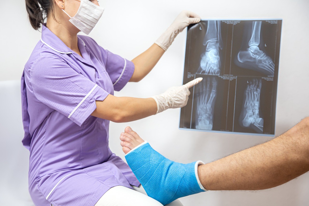
5. Fracture: The disruption of the bony cortex results in a fracture. Pain, edema, and deformity are brought on by the bleeding into the surrounding tissue from the broken bone.

a) Descriptive terms: Important details include the following: the affected bone, fracture location, whether the fracture is open or closed, whether the intra-articular extension has occurred, the type of fracture line, description of bone fragments (if present), whether the fracture is complete or incomplete, neurovascular status, and the position or alignment of the bone segments.

An open fracture occurs when the skin overlying the fracture is not intact either due to direct trauma or a fragment of bone that penetrates the skin. A subtle open fracture can occur when a bone fragment pierces the skin and then withdraws, leaving only a small puncture wound. Therefore, any fracture with an overlying skin wound should be suspected of being open. A closed fracture occurs when the skin and soft tissue overlying the fracture are intact.

An intra-articular fracture is a fracture that extends into the joint surfaces.

The fracture line may be spiral, oblique, or transverse. Fractures in children are often described as the buckle (torus), greenstick, or complete fractures. A fracture can also be described as compression, avulsion (chip), or comminuted (shattered) fracture.

The bone segments may be in contact with each other or separated by a measurable distance (usually stated in millimeters). The bone segments may be completely displaced (i.e., lying next to each other instead of end to end), partially displaced (i.e., offset from each other by a measured amount), or non-displaced.

The angulation of the two bone segments is described by both the direction of the angle (e.g., radial, dorsal, anterior, lateral) and by the degree of the angle formed by the two bone segments.

b) Special fractures

Pathologic fractures occur when a relatively minor force is applied to the diseased or otherwise weakened bone.

Stress fractures are caused by the repetitive application of a minor force to a bone, usually a long bone in the lower extremities. Stress fractures are commonly seen in military personnel (march fracture) and athletes (e.g., joggers, dancers).

Salter-Harris fractures involve the epiphyseal plate (i.e., the growth plate) and are common in children.

Comments (0)

Search
Popular categories
Latest blogs
All you need to know about Syphilis
ETIOLOGY: Syphilis is caused by T. pallidum.INCIDENCE: Each year, there are 29,000 new cases of syphilis. This figure represents probably only 10% of actual cases.CLINICAL FEATURES1. Primary syphilis: After an average incubation period of approximately 3 weeks, a smooth, painless ulcer called a chancre appears at the site of primary inoculation. The chancre heals without treatment in approximately 3 to 6 weeks; at about the same time, a painless uni- or bilateral regional adenopathy develops.2. Secondary syphilis represents disseminated disease and occurs in all patients with untreated primary infection. The lesions of secondary syphilis are papulosquamous lesions that occur over the entire trunk, extremities, penis, and buttocks. Fever and weight loss occur in 70% of patients.3. Tertiary syphilis occurs at least 10 years after the primary infection in at least 30% to 35% of untreated patients. The two most important manifestations of tertiary syphilis are cardiovascular syphilis, causing thoracic aneurysms, and neurosyphilis, causing meningitis, stroke, seizures, dementia, general weakness, and posterior column dysfunction.DIFFERENTIAL DIAGNOSES— Chancroid— HSV type 1 infection— Lymphogranuloma venereum— Tinea, sarcoid— Lichen planus— Seborrhea dermatitis— Molluscum contagiosum— Traumatic ulcer— Furuncle— CarcinomaEVALUATION: The clinical diagnosis can be confirmed by darkfield microscopic examination or more commonly serologic testing.THERAPY1. The standard treatment for primary, secondary, and early tertiary syphilis is benzathine penicillin G (2.4 million U administered intramuscularly as a single dose).2. For late tertiary syphilis or neurosyphilis, benzathine penicillin G (2.4 million U, three doses administered intramuscularly 1 week apart) is used. Doxycycline (100 mg orally twice daily for 14 days) can be given to patients who are allergic to penicillin.DISPOSITION1. Primary and secondary syphilis can be treated on an outpatient basis.2. Patients with neurosyphilis or major cardiovascular manifestations require admission for intravenous therapy.

Tue, 15 Nov 2022

What is Pemphigus Vulgaris?
Pemphigus Vulgaris is a rare disease that affects elderly patients. The mortality rate is 10%; most deaths result from steroid complications, secondary infection, dehydration, or thromboembolism. Pemphigus Vulgaris is caused by the attachment of immunoglobulin G autoantibodies to the epidermis. It has been associated with D -penicillamine and captopril administration.CLINICAL FEATURES1. Mucosal lesions and erosions are very common. Examination of all mucosal sites is warranted.2. Non-pruritic, painful, flaccid bullae appear that rupture easily. Blisters can be extended or new bullae formed by applying firm tangential pressure on the intact epidermis.3. Weakness, weight loss, and dysphagia may be presenting complaints.DIFFERENTIAL DIAGNOSES— Erythema multiforme— Bullous impetigo— Herpes zosterEVALUATION: Biopsy of lesions shows eosinophils, intraepidermal bullae, and acantholysis. Indirect immunofluorescent staining shows immunoglobulin G antibodies. Serum titers can be followed to evaluate the effectiveness of therapy.THERAPY1. Prednisone (200 to 350 mg/day) for 5 to 10 weeks is used until the cessation of new blister formation occurs. The dosage is then reduced to 40 mg on alternative days and tapered over 1 year.2. Azathioprine (100 mg/day) is added to the regimen and the dosage is reduced over a 4- to 6-month period. Methotrexate and cyclophosphamide can be used instead of azathioprine.3. Topical analgesics (e.g., viscous lidocaine) can be used to alleviate the pain associated with oral lesions.DISPOSITION: Patients with severe cases and oral lesions may require hospital admission for intravenous hydration. Others can be treated as outpatients with close follow-up.

Tue, 15 Nov 2022

Know about Scorpion Stings
Introduction: A nocturnal arachnid that lives in the Southwest of the United States is the scorpion. It has two venom glands and a stinger in its tail. The majority of species are rather benign, and they often only create a limited reaction similar to that brought on by a bee sting. The bark scorpion (Centruroides sculpturatus) venom, on the other hand, has neurotoxic that can result in a serious reaction. This dangerous scorpion is found on trees in Arizona and New Mexico.CLINICAL FEATURES1. Local effects: The C. sculpturatus scorpion bite causes immediate, excruciating pain at the stung site, as well as swelling and ultimately, numbness. The area that was hurt is extremely sensitive, and the implicated extremity could become paralyzed.2. Systemic effects: The neurotoxin is strongly cholinergic and can cause excessive salivation, blurred vision, muscular spasms, hypertension, and respiratory difficulties.DIFFERENTIAL DIAGNOSES — Snakebite— A puncture wound or other trauma— Insect or spider bite— Drug intoxicationEVALUATION: Typically, the offending scorpion is seen or assumed by history; if safety allows the scorpion to be brought in, this is best. Due to the wide range of symptoms and quick progression, a thorough history and physical examination are necessary.THERAPY1. Pre-hospital management includes rapid transportation of the patient, application of an ice pack to the sting site, and safe transport of the scorpion for identification. When serious symptoms appear, life-saving procedures should be started.2. ED managementa) Antivenin should be administered in all cases of severe envenomation.b) Ventilatory support may be required, with intubation and oxygen for patients with severe systemic response or anaphylaxis.c) Atropine may be required to counteract the cholinergic effects; the dose is titrated to relieve the cholinergic signs.d) Benzodiazepines may be used for seizures and muscle spasms.DISPOSITION: All victims should be observed for 24 hours, especially children. Symptomatic patients should be transferred to the intensive care unit if symptoms are severe.

Sat, 12 Nov 2022

All blogs