Published - Wed, 19 Oct 2022

Sinus Infection (Sinusitis): Causes, Symptoms, Evaluation & Treatment

Sinus Infection (Sinusitis): Causes, Symptoms, Evaluation & Treatment

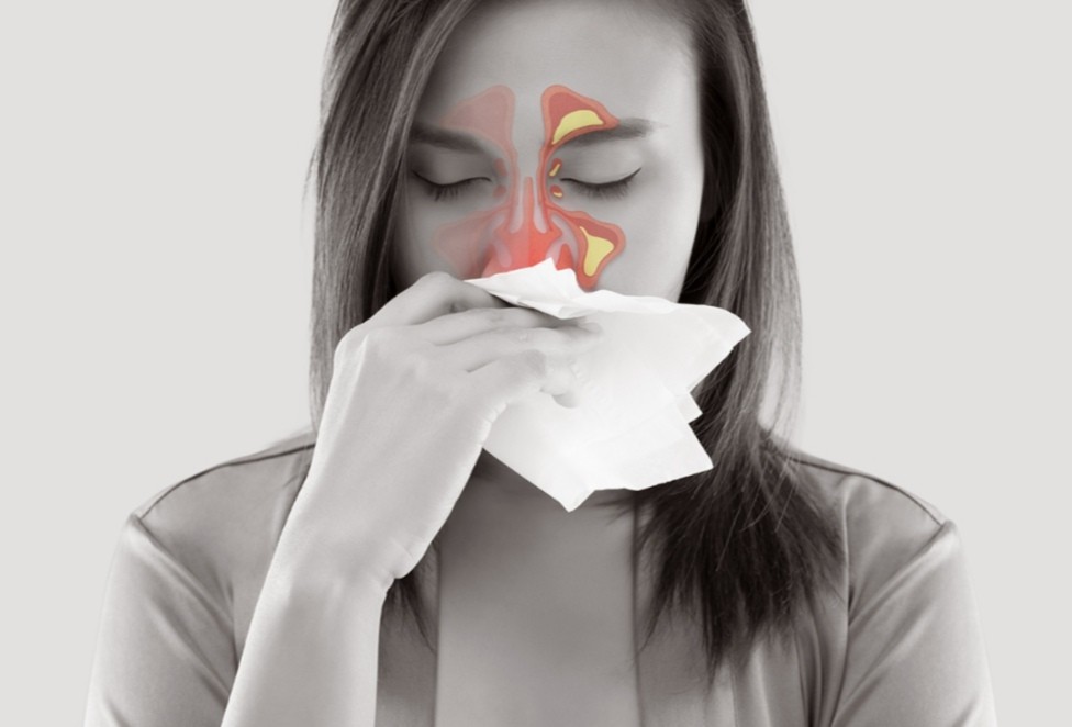
Sinusitis is an inflammation of the paranasal (ethmoid, frontal, maxillary, or sphenoid) sinuses. It is classified as acute or chronic, depending on how long the infection has been present.


ETIOLOGY AND PATHOGENESIS

Sinusitis occurs when pus is trapped in the sinus and is unable to drain completely through the Ostia into the nasal cavity.

a) Bacterial causes include H. influenzae, S. pneumoniae, streptococci, and M. catarrhalis.

b) Fungal causes are most common in patients with diabetes. Mucormycosis is particularly life-threatening.


PREDISPOSING FACTORS

— Chronic nasal edema

— Nasal polyps

— Nasal allergies

— Preceding upper respiratory tract infection

— Upper tooth abscess


CLINICAL FEATURES

1. Symptoms

a) Pain: Nasal congestion increases the pressure in the sinus, leading to pain that often worsens when the patient bends over. Patients may complain of a headache. Facial pain often corresponds to the affected sinus:

— Cheeks and upper teeth—maxillary sinus

— Eyebrow area—frontal sinus

— Eyes or behind the eyes—ethmoid sinus

b) Generalized malaise and periorbital edema are common complaints.

c) Nasal discharge is often blood-tinged and purulent. A postnasal drip may cause coughing and a sore throat.

2. Physical examination findings

a) Fever is often present in acute sinusitis but is rare in chronic sinusitis.

b) Tenderness to percussion may be present over the affected sinus.

c) The opacity of the affected sinus may be evident during transillumination. A negative transillumination does not rule out sinusitis because the test is only positive when the sinusitis is advanced.


DIFFERENTIAL DIAGNOSES

— Viral rhinitis

— Allergic rhinitis

— Vasomotor rhinitis

— Tumors

— Foreign bodies

— Wegener granulomatosis


EVALUATION

1. Laboratory studies: Typically not indicated. In acute sinusitis, a CBC may reveal an elevated white blood cell count.

2. Imaging studies

a) Radiographs: Plain sinus films may reveal air-fluid levels, cloudiness, or thickening of the sinus mucosa.

b) CT scans may identify an underlying predisposing anatomic condition (e.g., abnormally narrowed outlets, tumorous or cystic obstructions).


THERAPY

1. Supportive measures

a) An effort should be made to avoid air-borne irritants (e.g., smoke, pollen).

b) Although steam inhalation and environmental humidification are useful adjuncts, aggressive systemic rehydration is essential to mobilize and prevent reaccumulation of the thickened secretions.

c) Analgesics, decongestants, antihistamines, and steroids may also be helpful.

2. Antibiotic therapy: Not indicated in most cases. If determined necessary, acceptable regimens include:

a) Amoxicillin clavulanate (500 mg three times daily for 2 to 3 weeks)

b) Doxycycline (100 mg twice a day for 10 days)

3. Surgery: Surgical drainage of the sinus may be indicated if medical treatment fails. Surgery may also be indicated to correct underlying anatomic defects.


DISPOSITION

All patients diagnosed with sinusitis should be followed up until clinically cured.

1. Discharge: In the absence of complications (e.g., orbital cellulitis, osteomyelitis, septic cavernous thrombosis, contiguous spread to involve the nervous system), patients can be sent home with instructions to follow up in 1 to 2 days.

2. Admission is indicated for extremely ill patients, patients with orbital edema, and patients with nuchal rigidity or altered mental status.

Comments (0)

Search
Popular categories
Latest blogs
All you need to know about Syphilis
ETIOLOGY: Syphilis is caused by T. pallidum.INCIDENCE: Each year, there are 29,000 new cases of syphilis. This figure represents probably only 10% of actual cases.CLINICAL FEATURES1. Primary syphilis: After an average incubation period of approximately 3 weeks, a smooth, painless ulcer called a chancre appears at the site of primary inoculation. The chancre heals without treatment in approximately 3 to 6 weeks; at about the same time, a painless uni- or bilateral regional adenopathy develops.2. Secondary syphilis represents disseminated disease and occurs in all patients with untreated primary infection. The lesions of secondary syphilis are papulosquamous lesions that occur over the entire trunk, extremities, penis, and buttocks. Fever and weight loss occur in 70% of patients.3. Tertiary syphilis occurs at least 10 years after the primary infection in at least 30% to 35% of untreated patients. The two most important manifestations of tertiary syphilis are cardiovascular syphilis, causing thoracic aneurysms, and neurosyphilis, causing meningitis, stroke, seizures, dementia, general weakness, and posterior column dysfunction.DIFFERENTIAL DIAGNOSES— Chancroid— HSV type 1 infection— Lymphogranuloma venereum— Tinea, sarcoid— Lichen planus— Seborrhea dermatitis— Molluscum contagiosum— Traumatic ulcer— Furuncle— CarcinomaEVALUATION: The clinical diagnosis can be confirmed by darkfield microscopic examination or more commonly serologic testing.THERAPY1. The standard treatment for primary, secondary, and early tertiary syphilis is benzathine penicillin G (2.4 million U administered intramuscularly as a single dose).2. For late tertiary syphilis or neurosyphilis, benzathine penicillin G (2.4 million U, three doses administered intramuscularly 1 week apart) is used. Doxycycline (100 mg orally twice daily for 14 days) can be given to patients who are allergic to penicillin.DISPOSITION1. Primary and secondary syphilis can be treated on an outpatient basis.2. Patients with neurosyphilis or major cardiovascular manifestations require admission for intravenous therapy.

Tue, 15 Nov 2022

What is Pemphigus Vulgaris?
Pemphigus Vulgaris is a rare disease that affects elderly patients. The mortality rate is 10%; most deaths result from steroid complications, secondary infection, dehydration, or thromboembolism. Pemphigus Vulgaris is caused by the attachment of immunoglobulin G autoantibodies to the epidermis. It has been associated with D -penicillamine and captopril administration.CLINICAL FEATURES1. Mucosal lesions and erosions are very common. Examination of all mucosal sites is warranted.2. Non-pruritic, painful, flaccid bullae appear that rupture easily. Blisters can be extended or new bullae formed by applying firm tangential pressure on the intact epidermis.3. Weakness, weight loss, and dysphagia may be presenting complaints.DIFFERENTIAL DIAGNOSES— Erythema multiforme— Bullous impetigo— Herpes zosterEVALUATION: Biopsy of lesions shows eosinophils, intraepidermal bullae, and acantholysis. Indirect immunofluorescent staining shows immunoglobulin G antibodies. Serum titers can be followed to evaluate the effectiveness of therapy.THERAPY1. Prednisone (200 to 350 mg/day) for 5 to 10 weeks is used until the cessation of new blister formation occurs. The dosage is then reduced to 40 mg on alternative days and tapered over 1 year.2. Azathioprine (100 mg/day) is added to the regimen and the dosage is reduced over a 4- to 6-month period. Methotrexate and cyclophosphamide can be used instead of azathioprine.3. Topical analgesics (e.g., viscous lidocaine) can be used to alleviate the pain associated with oral lesions.DISPOSITION: Patients with severe cases and oral lesions may require hospital admission for intravenous hydration. Others can be treated as outpatients with close follow-up.

Tue, 15 Nov 2022

Know about Scorpion Stings
Introduction: A nocturnal arachnid that lives in the Southwest of the United States is the scorpion. It has two venom glands and a stinger in its tail. The majority of species are rather benign, and they often only create a limited reaction similar to that brought on by a bee sting. The bark scorpion (Centruroides sculpturatus) venom, on the other hand, has neurotoxic that can result in a serious reaction. This dangerous scorpion is found on trees in Arizona and New Mexico.CLINICAL FEATURES1. Local effects: The C. sculpturatus scorpion bite causes immediate, excruciating pain at the stung site, as well as swelling and ultimately, numbness. The area that was hurt is extremely sensitive, and the implicated extremity could become paralyzed.2. Systemic effects: The neurotoxin is strongly cholinergic and can cause excessive salivation, blurred vision, muscular spasms, hypertension, and respiratory difficulties.DIFFERENTIAL DIAGNOSES — Snakebite— A puncture wound or other trauma— Insect or spider bite— Drug intoxicationEVALUATION: Typically, the offending scorpion is seen or assumed by history; if safety allows the scorpion to be brought in, this is best. Due to the wide range of symptoms and quick progression, a thorough history and physical examination are necessary.THERAPY1. Pre-hospital management includes rapid transportation of the patient, application of an ice pack to the sting site, and safe transport of the scorpion for identification. When serious symptoms appear, life-saving procedures should be started.2. ED managementa) Antivenin should be administered in all cases of severe envenomation.b) Ventilatory support may be required, with intubation and oxygen for patients with severe systemic response or anaphylaxis.c) Atropine may be required to counteract the cholinergic effects; the dose is titrated to relieve the cholinergic signs.d) Benzodiazepines may be used for seizures and muscle spasms.DISPOSITION: All victims should be observed for 24 hours, especially children. Symptomatic patients should be transferred to the intensive care unit if symptoms are severe.

Sat, 12 Nov 2022

All blogs