Created by - Dr. Prashant Raj
How do regular screening and testing decrease the chance of Kidney Failure?
The delicate nature of the excretory organ is not often talked about and is usually unnoticed. The kidneys act as a filtration system that helps to free the body of poisons and waste products while maintaining fluid balance to support wellness and the proper functioning of the body. However, over time with age, there is a decline in the functioning of the kidneys. For most, it is attributed to natural aging except for others; it is an accelerated decline in functioning as a result of alternative underlying problems like diabetes and cardiovascular disease. Chronic unwellness like renal disorder, nephropathy, nephritis, and chronic kidney disease (CKD) is usually dangerous because the disease usually manifests itself at late stages.Kidney unwellness, which eventually ends up in kidney disease, is one of the foremost leading causes of death in the world. Many of us with kidney disease don't even discover it till it's in an advanced stage. Hence, there's a need for regular tests and screenings to take steps to forestall kidney disease. Knowing the health of one’s kidneys is the key to understanding a way to protect them. Early detection and timely management, will help prevent kidney function deterioration. Conditions like diabetes and cardiovascular disease square measure signals to additionally look out for deterioration of kidney function. The majority of all cases of CKD occur in diabetic and hypertensive patients as reported by multiple epidemiologic studies conducted over the years. Hence, it's becoming more necessary that people with diabetes and/or cardiovascular disease endure regular tests and screenings to make certain of their kidney health. Kidney screening includes 2 simple basic tests. The presence of protein in the urine is an early marker of excretory organ pathology along with the rise in Serum creatinineThese are straightforward tests that can be done regularly to know the status of kidney health. Other signs to look out for include · Swelling in the ankles or feet· Undue weakness, tiredness· Reduced urine output. Whereas these signs don't undoubtedly mean an individual has nephropathy, they must get this checked out to get a proper diagnosis. Doctors usually recommend urine protein and serum creatinine at 6-12 month intervals. The excretory organ may be a resilient organ and if pathology is detected early enough, it can be efficiently managed well in time with the correct medication and lifestyle changes.
More detailsPublished - Fri, 30 Sep 2022

Created by - Dr. Prashant Raj
Organic Brain Disorders and Psychosis
1. Organic brain disorders: Characterized by impaired orientation and cognitive brain functiona) Dementia: Typically older patients with progressive memory loss plus a decline in executive function, aphasia (speech), agnosia (use of objects), or apraxia (organization). They may be agitated due to the inability to understand their surroundings and often have an overlying component of psychosis or deliriumb) Delirium: Characterized by the rapid onset (hours to days) of impaired orientation and cognition. These patients may have a readily treatable and reversible condition (e.g., hypoglycemia).2. Psychosis: Typically characterized by abnormal thought patterns, often with intact cognition. These patients often can perform calculations, memorize items, or converse, but they have bizarre ideas and thoughts. For a diagnosis of psychosis, one should have hallucinations, delusions, catatonia, thought disorder, or social impairments. Psychosis is frequently a complication of one of the mental illnesses described below, but it can also develop from drug addiction or intoxication (e.g., methamphetamine abuse, alcohol abuse or withdrawal, and prescription drugs). Psychosis typically presents for the first time in a patient’s teens to mid-30s; patients presenting with psychosis at older ages should be highly suspicious of organic causes.a) Schizophrenia: Characterized by delusions and hallucinations and is the most common cause of psychosis. These patients may present to the emergency department (ED) in a flattened mood and withdrawn state (catatonia), or they may be violent, paranoid, and suspicious of healthcare workers. Antipsychotic medications are the mainstay of treatment, both emergently and on a chronic basis.b) Mania: Associated with bipolar disorder, wherein patients have cyclical mood swings that vary from depression to mania. Mania is characterized by elevated mood and energy. Acutely manic patients will exhibit pressured speech, agitation, grandiose delusions, and insomnia. Sedating neuroleptics are often needed in the emergent setting to control the patient.c) Depression: Patients may present with psychotic features, although this is rare. Delusions are the most common psychotic feature seen in depressed patients; these patients are not usually violent or agitated.EVALUATIONEDs should have a defined plan for dealing with violent or abusive patients. If the patient is threatening, evaluation and treatment should always take place with several people in the room.a) History and physical examination: An attempt should be made to obtain as much information about the patient’s condition as possible from relatives, friends, paramedics, and other health care workers. If possible, take a history or perform a physical examination before restraining or sedating the patient.b) Laboratory studies and other studies (e.g., radiography) should be guided by the history and physical examination findings. Patients with known psychiatric disorders or dementia may require minimal workup. Standard tests for these patients often include an ethanol level, drug screen, and basic blood work.THERAPYa) Restraint and sedation: The priority in dealing with patients with organic brain disorders or psychosis is ensuring the safety of both health care workers and the patient. There are various ways to do this.i) Environmental seclusion: Placement of the patient in a quiet, darkened room will often prevent escalation of agitation in mildly agitated patients.ii) Physical restraint: Violent and severely agitated patients may require physical restraints. At least five or six ED personnel must be present to restrain each of the patient’s limbs and his or her trunk in unison. Physical restraints are only justified if the patient is an imminent threat to himself or herself or others.iii) Chemical restraint: Sedation may be required if the patient remains agitated. Quick and safe sedatives to use include droperidol, ziprasidone, haloperidol, or lorazepam.—Patients on phencyclidine or methamphetamines may require substantial doses (e.g., 10 mg droperidol, 6 to 10 mg haloperidol, 10 to 15 mg lorazepam) before control is achieved.—Because some antipsychotics may lower the seizure threshold, the use of benzodiazepines may be more appropriate in certain circumstances (e.g., cocaine intoxication).—Patients with acute uncontrolled psychosis often require the rapid administration of antipsychotics to gain control.b) Glucose, oxygen, thiamine, naloxone, and flumazenil should be considered for the altered patient. These agents may rapidly correct the causes of coma, delirium, or psychosis. Note: Flumazenil should only be considered for the benzodiazepine-naive patient who has overdosed.DISPOSITION— Acutely psychotic patients usually need inpatient psychiatric care. An involuntary psychiatric hold may need to be invoked.— Patients with delirium should be admitted to the hospital unless a readily reversible or minor cause is found in the ED.— Many patients with drug or alcohol intoxication can be observed in the ED until they are appropriate for discharge.— Suicidal or homicidal thoughts should be ruled out before discharge.
More detailsPublished - Sat, 01 Oct 2022

Created by - Dr. Prashant Raj
Anorexia Nervosa and Bulimia Nervosa
Both eating disorders share similar symptoms but have characteristically different food-eating behaviors. Approximately 80% to 85% of patients are women younger than 25 years of age. Others are men or older women.1. Anorexia nervosa: Syndrome of self-starvation.2. Bulimia nervosa: Characterized by frequent episodes of binge eating. The patient usually consumes large amounts of food during a defined period (e.g., less than 2 hours) and then uses compensatory measures (e.g., laxatives, diuretics, induced vomiting) to prevent weight gain.CLINICAL FEATURES1. Anorexia nervosa: The patient usually weighs less than 85% of the expected weight (e.g., body mass index < 19).— The patient may present with life-threatening malnutrition, hypotension, or bradycardia.— Endocrine abnormalities are present, usually amenorrhea.2. Bulimia nervosa: These patients are often of normal weight or are overweight.— Serious complications of repeated vomiting (e.g., esophageal rupture, Mallory-Weiss tears, dental decay) may be the presenting complaint.— Electrolyte imbalances (e.g., hypokalemia) are fairly common secondary to repeated vomiting or laxative or diuretic abuse.DIFFERENTIAL DIAGNOSES1. Medical causes of excessive weight loss (e.g., diabetes, Crohn's disease, cancer, thyroid disorders, lupus) must be excluded.2. Anxiety, schizophrenia, body dysmorphic disorder, and depression can all lead to significant weight changes. Criteria for depressive disorder are found in nearly 50% of patients.EVALUATION1. History and physical examination form the basis for the diagnosis of these disorders— There is often a family history of eating disorders or job pressures to be thin (e.g., professional dancers, and gymnasts).— The patient is often evasive about purging or the use of laxatives or diuretics. Friends or family members are more likely to mention these behaviors.2. Laboratory studies: Appropriate studies include a serum electrolyte panel and glucose level as well as renal and thyroid function tests.3. Electrocardiography: An electrocardiogram is warranted especially in a symptomatic patient.THERAPYIntensive psychiatric therapy and frequent monitoring of weight are the cornerstones of treatment for patients with these disorders. Antidepressants are effective in certain patient populations.DISPOSITION1. Discharge: Most patients will require outpatient treatment.2. Admission to the hospital is indicated for any patient with substantial electrolyte imbalances or signs of cardiac abnormalities.
More detailsPublished - Sat, 01 Oct 2022

Created by - Dr. Prashant Raj
Hemoptysis- What it is, Causes & Treatment
Hemoptysis is the coughing /spitting up of blood from the respiratory tract. It's unremarkably caused by pneumonia, bronchiectasis, bronchitis, and carcinoma. The expectorated blood sometimes originates from the vessels. Once hemoptysis is observed then it should be classified based on severity, and the origin and reason behind the hemoptysis is determined. Lateral and AP chest X-ray is the initial study, though a standard chest X-ray doesn't rule out the chance of malignancy or any underlying pathology.Multidetector CT (MDCT) should be performed in patients with frank hemoptysis, suspicion of bronchiectasis or associated risk factors for carcinoma, and in those with signs of pathology on chest X-ray. MDCT may be a non-invasive imaging technique that will pinpoint the presence, origin, variety, and source of the general (bronchial and non-bronchial) and pneumonic blood vessel sources of bleeding. Endovascular embolization is the safest and simplest methodology for managing bleeding in severe hemoptysis. Flexible bronchoscopy plays a crucial role in the diagnosing of hemoptysis in patients with frank hemoptysis. The procedure is performed in the intensive care unit; it is used for immediate management of bleeding and is additionally effective in locating the source of the hemorrhage.Causes of hemoptysis -There is a broad medical diagnosis for hemoptysis, associated with infections, tracheal involvement, malignancy, and foreign body aspiration or trauma. In developing countries, one of the foremost common causes of hemoptysis is TB. However, in developed countries, bacterial infection superimposed on chronic respiratory sickness, like Cystic Fibrosis is also the cause. Acute infectious respiratory disease, pneumonia; and other infectious agents, like Aspergillus, Nocardia, and non-tuberculosis mycobacteria, may additionally be accountable.Finally, bleeding disorders, medicine use, foreign body aspiration, and respiratory organ trauma can contribute to the etiology of hemoptysis.Treatment for hemoptysis –Treatment of hemoptysis depends on the underlying cause and severity. In large hemoptysis or severe hemoptysis, securing the patient’s airway is a priority. Start on Basic medical aid, intubated, or repositioned in the lateral attitude position (i.e., on the affected side), which uses gravity to stop blood from getting into the unaffected lung. Intravenous fluid and transfusion are also indicated. Bronchoscopy to insert a balloon tube or embolize the artery to prevent further bleeding can be used. In some circumstances, surgical intervention is also needed. In non-massive hemoptysis, management of the underlying cause is the first treatment strategy. Infection is treated with antibiotics, antivirals, or antifungals. Signs of malignancy might need a consultation with an oncologist. Underlying chronic respiratory sickness is also treated with glucocorticoids, bronchodilators, mucolytics, chest physiotherapy, or basic first aid. Studies have additionally shown that inhalation of tranexamic acid (i.e., antifibrinolytic) in people with mild hemoptysis provides fast symptomatic relief and shorter hospital stays.
More detailsPublished - Mon, 03 Oct 2022

Created by - Dr. Prashant Raj
Minor heat illness
Most cases of minor heat illness occur within the first several days of exposure to a hot environment.CLINICAL FEATURES1. Heat cramps are brief, often severe muscular cramps that typically affect muscles heavily used by workers or athletes who are sweating profusely in hot environments. The cramps usually occur after the activity has ceased, while the person is relaxing, possibly resulting from salt deficiency (after sweating).2. Heat syncope is related to the vasodilation that occurs in people (particularly the elderly) exposed to hot conditions. The dilatation of cutaneous arteries causes a drop in cardiac output, relative hypovolemia of the thoracic blood vessels, a reduction in central venous return, and a reduction in brain perfusion. The dehydrated person is at high risk for syncope.DIFFERENTIAL DIAGNOSESIn cases of heat edema, it's important to rule out congestive heart failure, deep vein thrombosis, and lymphedema. Other significant causes of syncope should be ruled out because it might have a wide range of causes.EVALUATION1. An accurate history should be obtained, including the length of exposure to heat, type of work or activity, intake of water or food, salt intake, and events surrounding the onset of symptoms.2. Studies: In patients with severe heat cramps or syncope, analysis of serum electrolytes and a CBC may be required to guide therapy. ECG may be important as well in cases of injury.THERAPY1. Heat cramps are treated with rest and replacement of the deficient salt with an oral salt solution or may require intravenous isotonic saline (0.9% sodium chloride). Most patients respond rapidly to treatment.2. Heat syncope usually resolves when the person faints and assumes a supine position. Intravenous rehydration is frequently recommended because the patient may be dehydrated. People who are susceptible to heat syncope should be advised to take preventative steps like moving around regularly, flexing their legs when standing, and sitting or lying down anytime they experience early symptoms (e.g., vertigo, nausea, weakness) appear.DISPOSITION Minor heat diseases are easily treated, and most are prevented by patient education and providing proper fluid and salt replacement.
More detailsPublished - Tue, 04 Oct 2022

Created by - Dr. Prashant Raj
Home-cooked food increases your fitness efficiency
Eating home-cooked meals means that you are gonna adhere to DASH and Mediterranean diets, will be on bigger fruit and vegetable intakes and have better antioxidant consumption. Frequent consumption of home-cooked meals was related to a higher chance of getting a lower BMI and reduced body fat. Some studies reported that homecooked food is associated with normal HbA1c, and reduced cholesterol levels thereby preventing cardiovascular diseases in long run. Those who consume home medium meals 5 times, compared with 3 times per week, were 28% less overweight and had a less proportion of body fat In a big population-based cohort study, consumption of homecooked meals was related to higher dietary quality and lower fattiness. More prospective studies are needed to spot whether or not the consumption of homecooked meals has causative effects on diet and health.The prevalence of diet-related non-communicable diseases (NCDs), like type II Diabetes Mellitus, cardiovascular disease, and certain cancers, is increasing steadily worldwide. These changes are in the course of a decrease in the time spent on self-care, hugely relying on readymade food due to work-life imbalances, and changing lifestyles in the majority of developed countries. Concern has been expressed by health practitioners and researchers in the field of food and nutrition concerning a perceived decline in a change of lifestyle habits that have been hypothesized to be coupled to the rise in diet-related NCDs
More detailsPublished - Tue, 04 Oct 2022

Created by - Dr. Prashant Raj
Is Meditation best for depression?
If you're feeling a bit skeptical for no reason, you’re not alone. you'd probably even feel sadder when some people easily recommend that depression will improve if you merely “Smile more!” or “Think positively!”Depression can involve an excellent deal of dark thoughts. One may probably feel hopeless, worthless, or angry at life (or even himself) and meditation may seem somewhat unreasonable to a depressed person. But meditation teaches you to concentrate on thoughts and feelings while not passing judgment or criticizing yourself. Moreover, it involves increasing awareness of thoughts and experiences.Meditation doesn’t involve pushing away these thoughts. Instead, it helps you to observe and accept them, then permit them to pass through your mind. In this way, meditation can disrupt cycles of negative thinking.Say you’re sharing a peaceful moment alongside your partner. You're feeling happy and pampered. Then the thought comes to your mind, “They’re about to leave you alone and go away,” and then this thought overpowers you and keeps bothering you. What should you do? Feeling hopeless and stressed?MEDITATION can surely help. It can assist you to -• Notice this thought• Observe the risks associated with the thought• Acknowledge that it’s not the only riskMeditation can make it easier to concentrate on your emotions as they're easily accessible, especially, when you start experiencing negative thought patterns or notice raised irritability, fatigue, or less interest in the things that you would normally love to do. This is the time one should choose to engage in self-care for things from getting worse.How shall I do it?Meditation can feel discouraging if you’ve never tried it before, but it’s fairly easy and comfortable, once you start practicing itThese simple steps can assist you to get started:1. Get comfyIt’s typically helpful to sit down in a comfortable position when you initiate learning meditation, but if you feel like standing up or lying down, that works, too.The keys to feeling comfortable and relaxed. Closing your eyes will also help.2. Begin alongside your breathTake slow, deep breaths through your nose for a few secondsPay attention to:• How you breathe in, that is inhalation…• How do you breathe out or exhale• Feel the sounds of your breathYour thoughts could take your focus away from your breath, and that’s pretty normal. Just keep redirecting your focus to the breathing process whenever you catch yourself puzzling over any other thought.3. Move from breath to bodyEventually, start shifting your attention from your breath to the various components of your body to perform what’s known as a body scan.Start your body scan wherever you want to start. Some people start with their feet, whereas others like to begin with their hands or head. Focus your awareness on your body, moving from one part to another as you continue to breathe slowly and deeply.
More detailsPublished - Tue, 04 Oct 2022

Created by - Dr. Prashant Raj
Heat Exhaustion
A clinical illness known as heat exhaustion is defined by volume depletion in patients who have been subjected to heat stress. The majority of heat exhaustion instances in people who are in hot environments are caused by combined salt and water depletion following insufficient fluid and salt supplementation.CLINICAL FEATURES: There are many different heat exhaustion indications and symptoms.1. Symptoms: Early complaints of fatigue and vague malaise may progress to weakness, vertigo, nausea and vomiting, and headache.2. Physical examination findings: With significant dehydration, signs may include muscle cramps, orthostatic syncope, tachycardia, hyperventilation, and hypotension. Body temperature is normal or slightly elevated. Sweating may be profuse. Mental function is unaffected, and there are no indications of significant CNS injury.DIFFERENTIAL DIAGNOSES It includes cerebrovascular accident, drug ingestion, exacerbation of preexisting medical illness, psychological factors, infection, viral syndromes, and heat stroke.EVALUATION1. A history and physical examination should lead to an accurate diagnosis.2. Laboratory studies: There is no need for laboratory tests in mild cases. A CBC, a serum electrolyte panel, and a liver function panel may be useful in diagnosing hypernatremia, hyponatremia, hemoconcentration, or hepatic injury in moderate- to severe-case situations. The degree of dehydration can be assessed using urine-specific gravity readings and blood urea nitrogen/creatinine ratios.THERAPY: The patient should receive aggressive treatment for potential heat stroke if there is any doubt regarding the severity of the heat illness.1. Cool environment: The patient with an elevated body temperature should be cooled using a room-temperature water mist spray and fan to aid in evaporation. Cool packs are placed on the neck, axilla, and groin speed cooling.2. Correction of volume and electrolyte imbalances: Usually, symptoms resolve rapidly with intravenous and oral hydration.— The patient's condition should dictate the kind and amount of fluid.— To avoid cerebral edema, the free water deficit in the hypernatremic patient should be gradually restored over 48 hours. Severely hyponatremic patients should also be treated carefully and rectified gradually.DISPOSITION1. Discharge: In young, healthy patients who respond rapidly to treatment, no additional testing is required; these patients may be discharged with education about preventive techniques.2. Admission: Older patients, particularly those with cardiovascular disease or serious illness, require more careful fluid and electrolyte replacement and may need admission. Extremely young patients may also need extended intravenous fluid therapy.
More detailsPublished - Tue, 04 Oct 2022

Created by - Dr. Prashant Raj
Clonidine Toxicity
Clonidine is used for the treatment of hypertension and as adjunctive therapy in the treatment of opiate withdrawal. It is available in oral and transdermal preparations.1. Pharmacokinetics: Clonidine is well absorbed from the gastrointestinal tract and widely distributed in the body. The blood-brain barrier can be penetrated by clonidine. Within an hour, the effects start to show.2. Mechanism of action: Clonidine is a centrally acting α2 agonist that decreases peripheral sympathetic stimulation, decreasing norepinephrine activity and lowering systemic vascular resistance, heart rate, and cardiac output.3. Pathophysiology: Hypotension induced by α2 agonist activity. The drug also possesses opiate-like CNS depressant activity.CLINICAL FEATURES1. Cardiovascular features include bradycardia and hypotension: Early paradoxical hypertension may occur transiently due to vasoconstriction initiated by alpha-1 adrenergic stimulation 2. Neurologic features include miosis, sedation, coma, hypotonia, hyporeflexia, respiratory depression, and apnea.DIFFERENTIAL DIAGNOSESConsider β blocker or calcium channel blocker overdose, narcotic or sedative-hypnotic overdose, stroke, head injury, and hypoglycemia.EVALUATION: An ECG, ABG determination, chest radiograph, glucose level, and serum electrolyte panel should be obtained.THERAPY1. Initial stabilization includes airway management, cardiovascular resuscitation, and continuous cardiac monitoring.a) Initial transient hypertensive symptoms may be treated with nitroprusside infusion.b) Hypotension may be treated with intravenous fluid boluses or dopamine infusion.c) Bradycardia may be treated with atropine.2. Gastric decontamination is done with activated charcoal.3. Antidote treatment: Naloxone may be of benefit in reversing CNS depression. It has been reported to alleviate respiratory toxicity to a certain degree.DISPOSITION1. Discharge: Patients who receive activated charcoal and are asymptomatic after 6 hours of observation may be discharged.2. Admission: Symptomatic patients must be admitted to a cardiac monitoring setting.
More detailsPublished - Wed, 05 Oct 2022
Search
Popular categories
Latest blogs

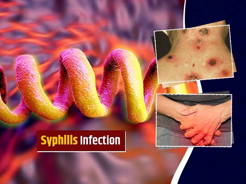
All you need to know about Syphilis
Tue, 15 Nov 2022

What is Pemphigus Vulgaris?
Tue, 15 Nov 2022

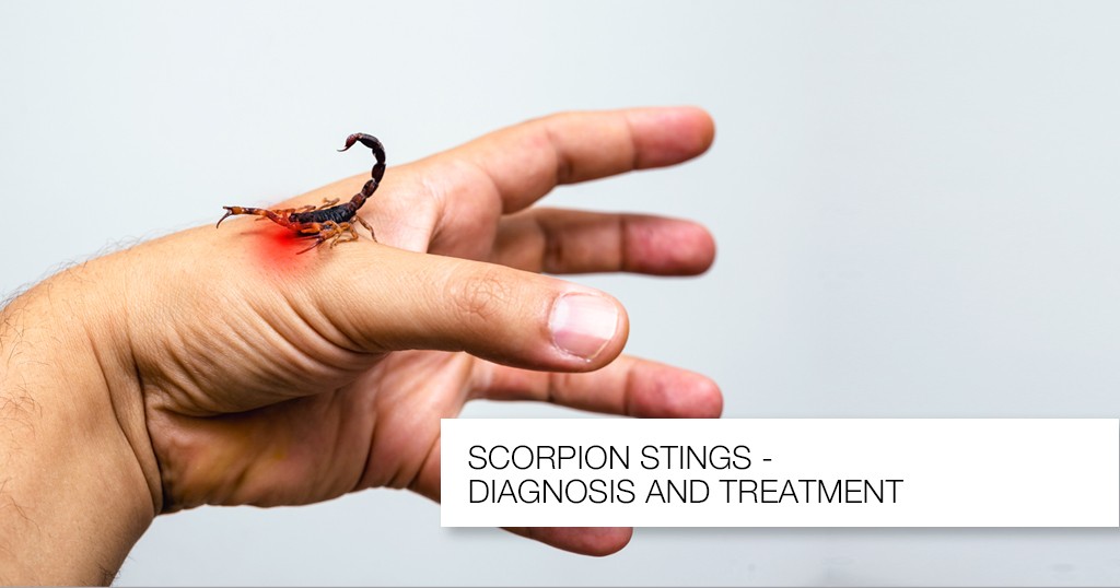
Know about Scorpion Stings
Sat, 12 Nov 2022
Write a public review