
Created by - Dr. Prashant Raj
PELVIC PAIN AND ITS CLINICAL FEATURES
There are many reasons for a female patient to have pelvic pain. There are primarily two categories of pelvic discomfort.1. Visceral pain —Results from stimulation of the nerves innervating the splanchnic organs. —The Character of pain: Pain is vague, dull, and poorly localized.2. Somatic pain —Results from stimulation of the peripheral somatic nerves. The pain is dermatomal or localized. —The character of Pain: Sharp pain typically affects the epidermis, a muscle, or the parietal peritoneum.CLINICAL FEATURES: When attempting to determine the source of the pain, it can be useful to be aware of the four different patterns of pelvic discomfort.1. Mild visceral pain results from visceral inflammation due to infection or distention. Causes include the following:a) Pelvic inflammatory disease (PID) is characterized by vague lower abdominal pain and diffuses cervical, uterine, and adnexal tenderness.b) Early appendicitis is characterized by diffuse abdominal pain, primarily epigastric or periumbilical, with right lower quadrant tenderness on direct palpation. Symptoms include fever, nausea, and anorexia, usually evolving over 24 to 48 hours.c) Vaginitis or cervicitis can cause diffuse, lower abdominal pain focused around the suprapubic region. Tenderness is appreciated on vaginal and cervical examination with no adnexal findings. Other symptoms include vaginal discharge, dyspareunia, and dysuria.d) Urinary tract infection is characterized by suprapubic fullness and crampy pain, possibly referred to the flanks. Patients may describe dysuria or hematuria associated with painful, burning urethral irritation.e) Ovarian cyst symptoms usually occur around midcycle or during the postovulatory phase. Fluid engorgement and distention cause dull, constant, achy pain in the lower abdominal adnexal region. Adnexal tenderness may be noted around the affected ovary.f) Early ectopic pregnancy: Distention at the site of implantation, nebulous lower abdomen pain, and/or adnexal tenderness are symptoms brought on by the expanding trophoblast. Vaginal bleeding and a positive pregnancy test are further results.g) Menstrual cramps are vague, cramping, sometimes sharp pains, lasting several days before to several days after the cycle begins. This is a diagnosis of exclusion.h) Uterine fibroids are associated with pubic pressure, pain, dysmenorrhea, or menorrhagia. Uterine tenderness may be noted, and fibroids may be palpable.i) Endometriosis: Ectopic endometrial tissue growth can result in a dull, ongoing pain that gets worse at the end of the menstrual cycle as a result of hormonal changes and tissue breakdown. As the lesions "slough," creating local inflammation, pain may become more intense during menstruation. Without cervical motion tenderness, the exam may detect adnexal or cul-de-sac tenderness. In the emergency room (ED), endometriosis is a diagnosis of exclusion that is often made only after laparoscopic surgery.j) Dysmenorrhea is cyclic, painful menstruation thought to be mediated by prostaglandins. The patient may describe a dull, progressive pelvic ache that radiates to her back or thighs.2. Severe visceral pain is an intense, restless pain often associated with nausea and vomiting. Underlying causes include severe infection, inflammation, obstruction, and visceral ischemia.a) Ovarian torsion is characterized by sudden, severe pain that is out of proportion to the concomitant adnexal tenderness. Symptoms usually occur around midcycle and can also occur in pregnant patients. The patient may have a history of ovarian cysts.b) Nephrolithiasis is characterized by colicky, intermittent, restless pain that usually begins in the flank but may progress or radiate to the lower quadrant, suprapubic, and groin regions. Associated nausea, vomiting, and hematuria are possible. The examination may reveal flank and/or lower quadrant tenderness without peritoneal signs.c) Bowel obstruction is associated with diffuse, crampy pain, bloating, nausea, and vomiting. An examination may reveal a distended abdomen, decreased or high-pitched bowel sounds, and diffuse tenderness without peritoneal signs. Inability to pass stool or flatus should alert the physician to this diagnosis. Previous abdominal surgery, hernia, or cancer are risk factors.3. Visceral pain progressing to somatic pain: Progressive irritation of the nearby peritoneum results from organ inflammation. Localized, somatic pain may be caused by an irritated parietal peritoneum.a)Appendicitis: Parietal peritoneal inflammation may be brought on by the rupture or progression of appendiceal inflammation. The patient reports generalized, crampy pain that gradually narrows in on the right lower quadrant. There is a sharper, more powerful, and more concentrated tenderness there. While appendicitis still occurs during pregnancy, the position of the pain and tenderness may change as a result of uterine displacement.b) Complicated PID: Local peritonitis can result from salpingeal infection spreading to the parietal peritoneum. As a result of diaphragmatic irritation, gonococcal or chlamydial seeding of the Glisson capsule (in the liver) can cause significant right upper quadrant discomfort and tenderness as well as right pleuritic chest pain (Fitz-Hugh-Curtis syndrome).c) Advanced ectopic pregnancy: Hemorrhage can happen as trophoblastic tissue erodes the implantation site, leading to diffuse peritonitis, shock, progressive adnexal and cul-de-sac soreness, abdominal distention, and local peritoneal pain.4. Sudden somatic pain: Diffuse peritonitis can develop from parietal peritoneal inflammation brought on by blood, cystic fluid, pus, urine, or faeces. This inflammation can cause excruciating local abdominal discomfort.a) Ruptured ectopic pregnancy: Small quantities of bleeding can cause localised, abrupt, intense stomach discomfort and tenderness. The peritoneum becomes diffusely implicated as the bleeding progresses, leading to substantial peritoneal abnormalities during an examination.b) Ruptured ovarian cyst: Cyst rupture causes fluid or blood to leak onto the parietal peritoneum next to it, resulting in abrupt, severe, unilateral, and potentially diffuse adnexal discomfort and tenderness.— Follicular cysts rupture midcycle; rupture is associated with ovulatory pain (mittelschmerz).— Luteal cysts rupture late in the menstrual cycle and may be associated with significant hemorrhage and shock.
More detailsPublished - Sat, 30 Jul 2022

Created by - Dr. Prashant Raj
HOW TO EVALUATE PELVIC PAIN
1.Stabilization: Airway, respiratory, and cardiovascular status should be assessed, and shock should be treated immediately before making a diagnosis.2. Patient history: It is imperative to obtain a menstrual history in all women of childbearing age.3. Physical examination: Take into account the kind of pain and tenderness the patient is feeling. Does vaginal discharge, stomach bloating, or fever have an association? Asking the patient to cough, gently moving the bed, or using percussion is the best way to demonstrate peritonitis.4. Laboratory studiesa) Pregnancy test: Pregnancy tests should be administered to all women of childbearing age. This can aid in excluding ectopic pregnancy or miscarriage but cannot do so entirely.b) Blood work: A blood type and screen and a complete blood count (CBC) should be obtained.c) Urinalysis should be performed on a sample obtained by catheterization.d) Culture: Swabs from the cervical and vaginal areas are submitted for potassium hydroxide, wet prep, and culture as necessary. Patients who have a fever may want to explore blood and urine cultures.5. Other diagnostic toolsa) Abdominal and pelvic ultrasonography may be indicated to search for free fluid in the cul-de-sac, ectopic pregnancy, ovarian cyst, ovarian torsion, uterine fibroids, tubal-ovarian abscess, or an inflamed appendix.b) Computed tomography scanning can be considered in the non-pregnant patient where ultrasound is unable to definitively identify the cause.c) Laparoscopy and biopsy may be necessary to definitively diagnose endometriosis, dysfunctional uterine bleeding, uterine or ovarian cancer, and complicated PID.
More detailsPublished - Tue, 02 Aug 2022

Created by - Dr. Prashant Raj
TREATMENT OF PELVIC PAIN
1. Vaginitis: a) Infectious vaginitis:i) Fungal vaginitis— C. albicans vaginitis is treated with miconazole suppositories (200mg each evening for 3 to 7 days), oral fluconazole (150 mg orally once), or terconazole suppositories (80 mg each evening for 3 days).— Other fungal agents of vaginitis (e.g., Torulopsis glabrata) may be resistant to conventional antifungal treatment and may need longer therapy, repeated dosing, or systemic antifungal treatment.ii) Trichomoniasis is treated with oral metronidazole (2 g administered in a single dose). In pregnant patients, a 7-day course of metronidazole (500 mg twice daily) or clotrimazole suppositories (inserted each evening for 7 days) can be used.iii) Bacterial vaginitis is treated similarly to trichomoniasis. Metronidazole (500 mg twice daily for 7 days). Treatment with clotrimazole suppositories can be considered for pregnant patients with severe infections.b) Foreign object vaginitis: Object removal and proper hygiene education.c) Contact vaginitis is treated by terminating exposure to the offending agent.d) Atrophic vaginitis is improved by applying topical estrogen cream daily for 5 to 10 days.2. Cervicitis: Gonococcal and chlamydial infections are the focus of treatment for bacterial infection due to high frequencies of coinfection.a) Gonococcal: Ceftriaxone (250 mg in one intramuscular dose) plus doxycycline (100 mg orally every 12 hours for 7 days). Doxycycline and tetracyclines should be avoided in pregnant women.b) Chlamydial: Azithromycin (1 g administered in a single oral dose) or a 7-day course of doxycycline.c) Viral: Herpes virus develops a latent state inside neuronal cells and cannot be removed from the body. To reduce the severity of the initial and reactivation symptoms, doctors may prescribe acyclovir (200 mg orally five times daily, or 400 mg orally three times daily for 10 days) or valacyclovir (1 g orally twice daily for 10 days).3. PID: a) Outpatient therapy: Ceftriaxone (250 mg administered in a single, intramuscular injection) plus doxycycline and metronidazole for 14 days. b) Inpatient therapy: Appropriate regimens include:—A cephalosporin (e.g., ceftriaxone, 1 g every 24 hours; cefotetan, 2g every 12 hours; or cefoxitin, 2 g every 6 hours) plus doxycycline (by mouth or IV).—Clindamycin (900 mg every 8 hours) plus gentamicin (an initial loading dose of 2 mg/kg followed by a maintenance infusion of 1.5 mg/kg every 8 hours intravenously).—Ampicillin/sulbactam (3 g IV four times daily) plus doxycycline.4. Ectopic pregnancy:a) General therapy: To avoid the possibility of maternal Rh antibody development, Rho(D) immune globulin is administered as a single intramuscular injection to all Rh-negative mothers. Possibly within 72 hours following the first incident of bleeding.b) Specific therapyi) Unruptured ectopic pregnancy: These patients are hemodynamically stable. Treatment options include:—Laparoscopic salpingostomy and ectopic excision.—Nonsurgical treatment with methotrexate, leucovorin, or mifepristone.—Salpingocentesis (i.e., transvaginal needle aspiration of the gestational sac, potentially with direct injection of methotrexate)ii) Ectopic pregnancy without cardiac activity: The obstetrician may choose to treat patients without surgery if their β-hCG levels have been serially declining and repeat sonography has revealed no signs of erosion or rupture. It is possible to induce evacuation using medication. If there is any indication of erosion or rupture, laparoscopic intervention is carried out.c) Ruptured ectopic pregnancy: These patients may be hemodynamically unstable, necessitating prompt fluid and blood resuscitation as well as bruising control. Salpingectomy with laparoscopic ectopic excision is advised.5. Ovarian cyst: If sonographic evidence of free fluid in the cul-de-sac is seen without the need for treatment, a patient with a confirmed cyst may be sent home with pain medication. Surgery should be performed on patients who show signs of bleeding that may be active or have hemodynamic instability.6. Ovarian torsion: Patients should have a laparoscopy or laparotomy for exploration and attempted salvage if their history, physical exam, and sonographic results are suspicious.7. Endometriosis: To stop ectopic endometrial growth, oral contraceptive methods, methyltestosterone, or progesterone may be utilized. In refractory cases, surgery is used to remove the tumor.8. Dysmenorrhea: It is possible to try oral contraceptive medication to slow endometrial development. Nonsteroidal anti-inflammatory medicines, such as ibuprofen or naproxen, are used to try to control pain. Drugs like opiates are usually avoided.DISPOSITION—Hemodynamically unstable patients are treated with aggressive fluid resuscitation (with blood products) and taken to the operating room.—Hemodynamically stable patients are admitted for observation and treatment when an evolving surgical disease is suspected or the patient is at risk for systemic toxicity.—All patients who are discharged must-have follow-up arranged and verified before leaving the ED.
More detailsPublished - Tue, 02 Aug 2022
Created by - Dr. Prashant Raj
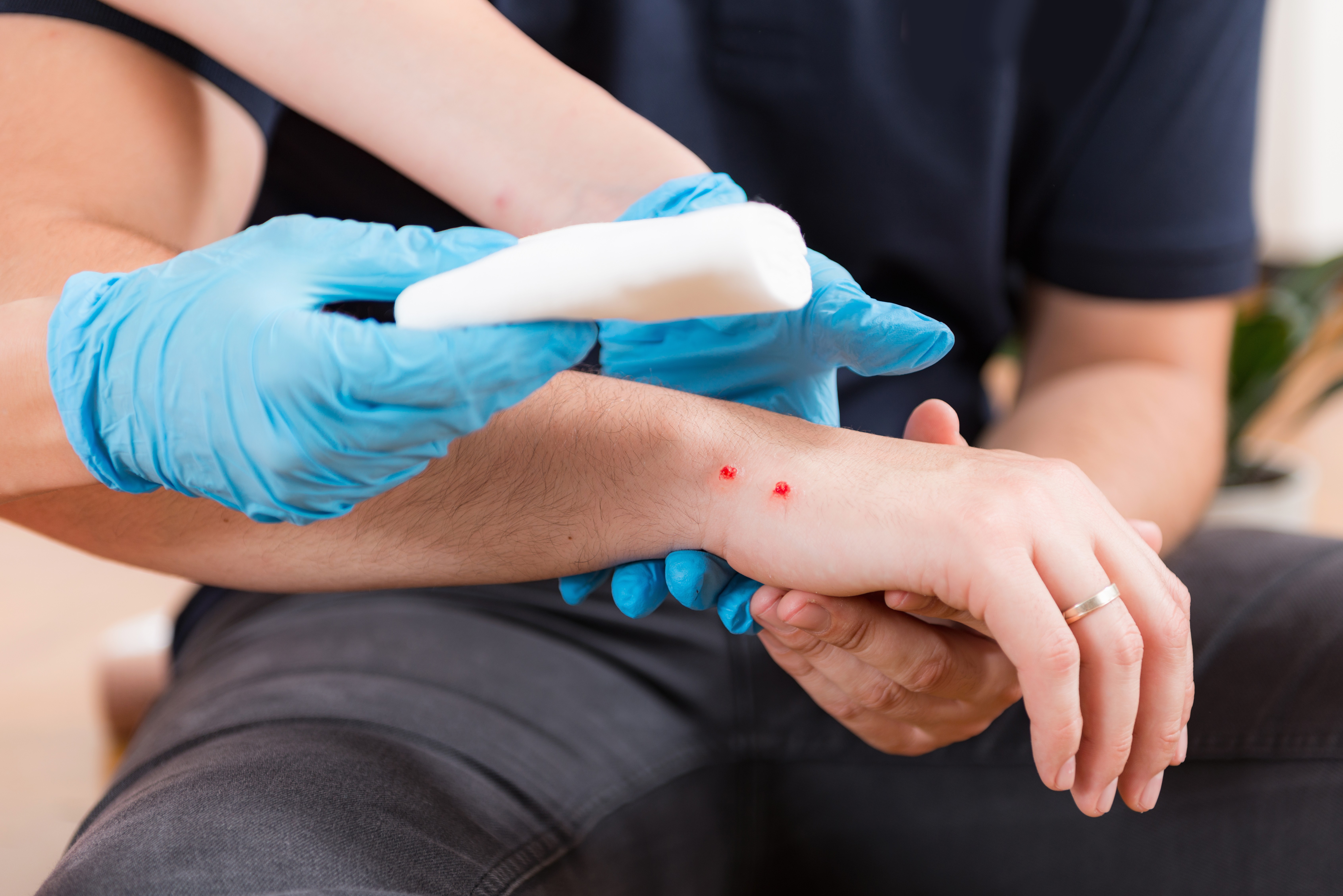
DO’s and Don’ts during Snake Bite
Today there are thousands of venomous animals thriving around the world and over time their venoms have evolved to do specific jobs envenomate their prey.Discover what venom is, why some species have implausibly potent venom, and why it is important to take immediate action after being bitten….There are many different less common uses for venom. For instance, male platypuses use their venomous spurs against their competition during the breeding season; chromatic crazy use theirs to counter the venom of fireplace ants and a few species, like shrews, are thought to use their venom to preserve food.Two types of venomous snakes are notably well known: vipers (Viperidae) and elapids (Elapidae). Broadly, the venoms in these 2 types affect the victim differentlyVipers, which include Adders and Rattlesnakes, have venoms that are typically haemotoxic which means the venom specifically attacks the vascular system and might interfere with the blood's ability to clot.Many venomous snakes are elapids, like cobras, mambas, kraits, and taipans. Their venom is usually Neurotoxin, which implies that it interferes with the transmission of nerve impulses. It typically results in either making a victim's body flip rigid or become limp.Symptoms of Snakebite• Redness, swelling, bruising, bleeding, or blistering around the bite• Puncture marks at the wound.• Severe pain and tenderness at the location of the bite• Nausea, or vomiting• Labored respiration (in extreme cases, respiration might stop altogether)• Rapid pulse rate, weak pulse, low blood pressure level• Disturbed vision•You feel the Metallic, mint, or rubber taste in the mouth• Increased sweating• You may feel numbness around your face and limbs.• Muscle crampFirst aid steps -Antivenom is the treatment for serious snake envenomation. The earlier antivenom is started, the less damage will be there in a patient Driving self to the hospital isn't suggested as individuals with snakebites have chances to become dizzy or may pass out.Do’s - • Take a photograph of the snake from a secure distance if doable. The identification of a distinctive snake will facilitate with correct treatment of the bite.• Keep calm.• Lay or sit down with the bite in a neutral position of comfort.• Quickly remove your watch and ring before swelling starts.• Wash the bite with soap and water.• Cover the bite area with a clean cloth and do the dry dressing at the bite area.• Mark the tenderness/swelling on the skin and write the time Don’t’s –• Do not catch the snake or try and lure it. Never handle a venomous snake, not even a dead one or its beheaded head.Do not await symptoms to look if bitten, get a medical emergency quickly.• Do not apply a bandage.• Do not slash the wound with a knife or cut it in any approach.• Don’t suck the venom from the mouth.• Don’t use ice or water around the venom area.• Do not drink alcohol as a medicinal drug.• Do not take pain relievers (such as Bayer, ibuprofen, naproxen).• Do not apply anything over the bite or take an electrical shock
More detailsPublished - Tue, 02 Aug 2022

Created by - Dr. Prashant Raj
HOW TO APPROACH ILL PEDIATRIC PATIENT
The emergency department (ED) sees between 30% and 35% of patients who are younger than 18 years old. Most of these kids can't get to the primary provider's office and need urgent treatment or office care for self-contained diseases. As a result, people come to the ED for treatment. True emergencies, in which children could die or suffer major disabilities if not treated right away, account for between 3 and 6 percent of ED visits.CLINICAL FEATURESExperienced pediatric emergency physicians frequently refer to children in urgent need of assessment as "septic" or "sick," as they quickly make the assessment based on a constellation of symptoms and indications that are challenging to teach without first-hand experience.Signs and Symptoms of a Sick Child— Lethargy with little or no apprehension to examination or painful procedures; failure to interact appropriately with their environment.— Drooling or stridor with severe air hunger.— Cyanosis of lips and extremities with poor perfusion and absent pulses in the lower extremities.— Extreme hypotension— Irritability, particularly when held by parents, with the inability to be comforted— Poor feeding is associated with a weak suck and poor interest in feeding— Elevated temperature or hypothermia.— Poor capillary refill with mottled skin appearance or poor turgor.— Persistent vomiting with or without feedings.— Complaint of headache and photophobia (in older children); the stiff neck is not a reliable sign in children younger than 18 months, and these children are unlikely to be able to report subjective complaints.— Seizures may be prolonged or focal and are often associated with fever.— Altered mental status, particularly with combativeness or inappropriate thoughts.— Respiratory distress, particularly with nasal flaring, grunting respirations, tachypnea, intercostal and subcostal retractions, or diaphragmatic breathing.— Any presentation of trauma that may be associated with a blunt injury to the head or thorax or with a penetration injury to the chest.— Petechiae or purpura associated with fever.— Weak cry or no cry when painful procedures or examination maneuvers are performed.DIFFERENTIAL DIAGNOSESChildren who suffer true, life-threatening emergencies fall into several distinct categories of illness. Children with any of the following life-threatening emergencies usually have initial respiratory compromise resulting from increased metabolic demands and cardiac compromise as a secondary event:— Acute respiratory distress— Cardiovascular disorders— Environmental injuries— Injuries and emergencies with altered states of consciousness— Shock syndromes— Traumatic disorders
More detailsPublished - Wed, 03 Aug 2022

Created by - Dr. Prashant Raj
Trauma - Types & Disorder
Trauma is the response to a deeply distressing or perturbing event that overwhelms the individual’s ability to cope and causes feelings of helplessness, diminishes their sense of self-worth, and their ability to feel emotions and experiences. While there aren't any objective criteria to gauge that events can cause post-trauma symptoms, circumstances usually involve the loss of management, betrayal, abuse of power, helplessness, pain, and confusion. The event needn't rise to the extent of war, or natural disaster, It could be a personal assault to have an effect on an individual deeply and alter their experiences. Traumatic things that cause post-trauma symptoms vary individually and quite dramatically from person to person. Indeed, it's subjective. Not each traumatized person develops post-traumatic stress disorder (PTSD). Some individuals develop some symptoms like those who are more sensitive, it might take some weeks to develop symptoms. This is often known as acute stress disorder (ASD). When the symptoms last over a month and seriously have an effect on the person’s ability to perform, the person could also be stricken by an anxiety disorder. Some individuals with anxiety disorder don’t show symptoms for months after the event itself. And a few individuals are affected by anxiety disorder symptoms of a traumatic experience for the remainder of their life. Symptoms of anxiety disorder will present as panic attacks, depression, self-destructive thoughts and feelings, drug abuse, feelings of being isolated, and not having the ability to finish daily tasks. There is no magic treatment that may heal you overnight, and neither is there one kind of psychotherapy that's right for everybody, however, it is advisable to seek treatment whenever you face any such issues. Healing is sort of a marathon. It needs preparation, recurrent observation, courage, determination, and also the support of others—including that of a health expert. When trying to find an expert, it's very important to keep in mind that, the medical aid is being offered as a collaborator and not being imposed on you. Additionally, it's crucial that you simply feel safe in your therapeutic relationship.Studies have found that people who actively participate in their medical aid gain more and get better quickly. Treatments offered for PTSD 1. Pharmacotherapy is the use of medicines to manage highly troubled trauma reactions. Medications are shown to be useful with the following reactions/symptoms: o Intrusive symptomso Hyperarousalo Emotional reactivityo Irritabilityo DepressionTaking medication doesn't mean trauma and associated pain will go after a single dose. Medications will slowly work till symptoms are less intense and manageable. If you choose to use medications, consult a shrink and continue consulting a shrink for as long as you're taking the medications. Inform the shrink of how the medications are impacting you. Some medications have a few side effects that might not be tolerable to you, and a few individuals don't respond favorably to medications. 2. BEHAVIOR medical aidThe most common type of behavior modification is exposure. In the desensitization technique, one is made to face one’s fears bit by bit. for example, the reminiscences of a traumatic event–without the dreaded consequence occurring. Often, this exposure ends up in the individual learning that the worry or negative feeling is unwarranted, which successively permits the worry to decrease. § Exposure therapy has been found to scale back anxiety and depression, improve social adjustment, and organize the trauma memory. There square measure varied kinds of exposure therapy: o Imaginal exposure: A person imagines the dreaded event as vividly as doable.o In vivo exposure: The exposure happens within the medical aid.o Systematic desensitization: The individual is exposed to in turn additional fear-inducing things. This exposure is paired with relaxation.Exposure therapy could be an extremely effective treatment for posttraumatic stress (PTSD). § Another kind of behavior modification is Stress immunization coaching (SIT), also called relaxation coaching. Stress immunization coaching teaches people to manage stress and anxiety. § COGNITIVE BEHAVIORAL THERAPY Cognitive Behavioral Therapy (CBT) is grounded within the concept a person should correct and alter incorrect thoughts and increase information and skills. Common parts of psychological feature activity include: o Teaching people a way to breathe to manage anxiety and stresso Educating people on traditional reactions to trauma Exposure therapyIdentifying and evaluating negative, incorrect, and irrational thoughts and substituting them with additional correct and fewer negative thoughts. § There is no guiding principle for psychotherapy. In general, a hypnotherapist guides the individual in medical aid into a hypnotic state, then engages the person in spoken communication or speaks to the person concerning bound key issues. Most hypnotherapists believe that the emotions and thoughts that a person comes into contact with whereas below mental state square measure crucial to healing. § PSYCHODYNAMIC THERAPYThe goal of psychodynamic trauma therapy is to identify which phase of the traumatic response the individual is stuck in. Once this is discerned, the therapist can determine which aspects of the traumatic event interfere with the processing and integration of the trauma. Common elements of psychodynamic therapy include: o Taking the individual’s developmental history and childhood into accounto Emphasizing on understanding the meaning of the traumao Looking at how the trauma has impacted the individual’s sense of self and relationships, as well as what has been lost due to the traumatic event § GROUP THERAPYThere are a variety of different groups for trauma survivors. Some groups are led by therapists, others by peers. Some are educational, some focus on giving support, and other groups are therapeutic. Groups are most effective when they occur in addition to individual therapy. A trauma survivor needs to choose a group that is in line with where one is in the healing journey: o Safety/victim phase: Choose a group focused on self-care and coping skills.o Remembering and mourning/survivor phase: Pick a group focused on telling the trauma story.o Reconnection/thriver phase: Join a group that aims to create a connection with people. o Educational groups are appropriate during all phases.
More detailsPublished - Thu, 04 Aug 2022

Created by - Dr. Prashant Raj
HOW TO EVALUATE ILL PEDIATRIC PATIENT
1. A primary survey should be performed immediately.a) Airway: The patency of the airway needs to be evaluated.— To remove tongue obstruction, the airway should be freed with external techniques.— Obstruction from foreign bodies should be recognized and relieved.— Gas exchange should be assessed and optimized.— Aspiration of gastric or oral contents should be prevented.— An artificial airway should be provided if no protective reflexes exist.b) Breathing: The adequacy of ventilation should be assessed, including the equality of chest rise and breath sounds.— A determination of whether air entry is impaired from a central or pulmonary cause should be made.— Ventilation should be enhanced if necessary with mouth-to-mouth breathing, bag-mask ventilation, or endotracheal intubation with or without bag assistance.— A surgical airway should be provided if an oral or endotracheal airway cannot be established.c) Circulation should be assessed, including the quality and intensity of pulses in the upper and lower extremity and blood pressure.— Hemorrhages should be controlled with direct pressure until surgical intervention can be accomplished.— Intravenous vascular access and a resuscitative fluid bolus should be provided; if intravenous access cannot be rapidly achieved, intraosseous access should be performed.— External cardiac massage should be provided if no pulse exists.— Pharmacologic management of circulation should be provided if the fluid status is adequate and cardiac output is impaired because of either vascular or pump instability.— Conversion of dangerous cardiac rhythm disturbances should be provided if hypotension and unresponsiveness ensue.d) Neurologic examination: A preliminary assessment should be made according to the following simple scale:— Alert and responsive to verbal stimuli— Responsive to verbal stimuli but no spontaneous alertness.— Responsive only to painful stimuli.— Unresponsive to any stimulie) Overall examination:— Infants should be unclothed and examined rapidly for injuries. — Passive heat loss should be prevented by removing wet clothing and by providing external warmth.— The patient’s core temperature should be obtained to monitor for hypo- or hyperthermia.2. A secondary survey, entailing a detailed physical examination and history, is then performed.a) Head— Evidence of trauma is determined by palpating bony prominences and the maxillae and by inspecting the nose and ears for drainage of cerebrospinal fluid (CSF).— The existence of dehydration is determined by inspecting the mucosae for evidence of decreased tearing or moisture or sunken eyes.— The eyes should be inspected for pupillary size and extraocular movements. A funduscopic examination should be performed if possible to assess for the central nervous system (CNS) or toxic involvement.— The oral cavity should be inspected for odor or discoloration that may imply a toxicologic basis for the condition.b) Neck— The neck should be palpated for midline tenderness or deformity.— Flexion, extension, and rotation should be performed only after an injury have been excluded either clinically or by radiographic means.c) Chest— The chest wall should be inspected for symmetric expansion or disarticulation.— Palpation for change in respiratory fremitus or inequality should be performed.— Auscultation of the chest for breath sounds and evidence of adventitial sounds such as rales, rhonchi, or wheezes should be performed.— Penetrating or blunt chest trauma is treated immediately.d) Abdomen— The abdominal wall should be inspected for bruising, penetration, or hematoma.— Palpation for trauma, masses, or guarding and rigidity, which may indicate peritonitis, should be performed.— Palpation of the flanks to search for hematoma or an expanding mass should be performed.e) Pelvis— The pelvis should be palpated laterally and in an anteroposterior position for tenderness or instability.— The perineum should be inspected for lacerations, active bleeding, or hematoma. The urethra and rectum should be inspected for trauma or blood.— In age-appropriate females, a pelvic and rectal examination should be performed, and pregnancy should be excluded.f) Extremities— The extremities should be inspected for signs of abrasion, hematoma, laceration, or deformity.— Palpation of the bones for instability, tenderness, or deformity should be performed.g) Neurologic assessment— Attention should be focused on cranial nerve function, motor function, and sensory function, along with the patient’s level of consciousness.— The presence or absence of reflexes should be noted.— Serial examinations for changes in the level of consciousness or loss of neurologic function should be performed.3. Additional studies are often nonspecific but may provide clues to the diagnosis.a) Laboratory studies— Aerobic and anaerobic cultures of blood— Arterial blood gas (ABG) determination— Complete blood count (CBC) and differential— Electrolytes and bicarbonate— Blood urea nitrogen and creatinine— Immediate fingerstick glucose and serum glucose— Urinalysis and urine culture— Stool cultureb) Radiologic studies— Abdominal radiography— Abdominal ultrasonography— Chest radiography— Computed tomography (CT) of the head/abdomenc) Ancillary studies— Echocardiography— Electrocardiography— Lumbar puncture— Urine for toxicologic analysis
More detailsPublished - Thu, 04 Aug 2022

Created by - Dr. Prashant Raj
Trauma - Reason for Death
The first peak within the classic trimodal model of trauma mortality is immediate death occurring within minutes of the injury. These patients are declared dead on the scene or die shortly when arriving at the hospital. In most revealed reports, these embrace deaths at the scene, deaths occurring within one hour of arrival to the hospital. These deaths are usually a consequence of severe injuries. The seminal works of Baker et al and Trunkey in the Seventies showed that 64% of trauma deaths occurred on the scene, with the patients not even transported to a hospital. Despite all the progress in emergency medical services and trauma systems, pre-hospital care, injury interference, and automotive safety, the proportion of deaths occurring in real-time when the injury has remained unchanged over time. The second peak within the trimodal distribution is early deaths, outlined as deaths within hours of arrival to the hospital. In most revealed reports, early deaths embrace deaths within twenty-four hours of arrival to a trauma center, excluding immediate deaths. These deaths also are a consequence of severe injuries; however, the patients reach the hospital alive and the injury is probably treatable with prompt definitive care.Deaths among trauma patients after discharge have gone unnoticed. This can be due to the issue of follow-up within the trauma patient population. First, the patient stays far from a trauma center. Second, trauma patients are usually younger people who stay away from home for education or work. Many studies have shown that trauma patients have a magnified risk of mortality when discharged. CAUSES OF DEATH DUE TO TRAUMA Several studies have investigated the reason for death in trauma patients. It was found that brain injury accounted for a majority of deaths [50%], Heart or aortal injury (17%), hemorrhage (12%), infection (10%), respiratory organ injury (6%), burn (3%), and liver injury (2%) accounted for the rest. The majority of patients with an internal organ, vascular, or liver injury died of hemorrhage. Figure 1: Cause of death in trauma patients Head injury was the foremost common cause of death. Almost a 3rd (31%) of victims died of hemorrhage within the chest, the abdomen, or inside the organ. Lack of pre-hospital care and shorter transport times to trauma centers remains the reasons for death in a grievous injury.
More detailsPublished - Fri, 05 Aug 2022

Created by - Dr. Prashant Raj
TREATMENT & DISPOSITION OF ILL PEDIATRIC PATIENT
1. Stabilization is the priority: Before admission or transport, all life-threatening conditions (in the particular, tenuous airway or circulatory dysfunction) must be addressed fully.a) Before admission or transfer, any potential airway compromise must be treated, stabilized, and, if necessary, endotracheal intubation must be obtained.b) Complete attention must be given to the circulatory system; resuscitative fluid boluses or blood transfusions may be necessary.c) Vasoactive drugs (e.g., dopamine, dobutamine) should be administered only in the presence of a physician or a nursing staff qualified in critical care. The environment should be monitored and should have intravenous pump capabilities.2. Supportive care is essential:a) Fluid resuscitation: A fluid bolus of 20 mL/kg should be provided to establish blood volume and increase the efficiency of the heart. Care must be taken to avoid cerebral edema of fluid overload when the vascular volume is restored.b) Anticonvulsant therapy is indicated to control seizures.c) Assisted ventilation may be necessary to control respiratory failure or cerebral edema.d) Surgical consultation should be obtained promptly in all cases of suspected trauma or when the diagnosis is in doubt in a critically ill infant with nonspecific physical signs.3. Antibiotic therapy should be initiated for children with presumed sepsis, meningitis, or pneumonia.a) Agent selection: The patient should be treated with an antibiotic that is effective against the pathogens that most often cause disease in the patient’s age group.— Neonates and infants younger than 2 months are usually given ampicillin sodium and either an aminoglycoside or cefotaxime.— Infants and older children may be treated with ceftriaxone in single or daily divided doses or cefotaxime. Vancomycin should be added in unstable patients to cover for resistant treptococcus pneumoniae and methicillin-resistant Staphylococcus aureus.b) The duration of therapy is based on the patient’s age and the causative organism.— Infants with sepsis but without meningitis are usually treated with intravenous antibiotics for 7 to 10 days.— Infants with meningitis receive 10 to 14 days of parenteral therapy depending on the causative organism. Meningitis caused by Neisseria meningitidis may be treated for fewer than 7 days under certain circumstances.4. Corticosteroid therapy is indicated for patients with meningitis to reduce the incidence of hearing loss. These drugs also may augment survival in children with sepsis or meningitis, but the effective dosage is unknown.DISPOSITION1. Critically ill infants must be evaluated and treated promptly and admitted to the hospital for further treatment.a) Such children should be cared for by the highest level of provider available, preferably a pediatrician able to care for critically ill infants.b) When higher level care is unavailable, transport should be arranged to a referral center with the capability to care for critically ill children.2. Seriously ill infants who may have the possibility of a bacterial process should not have antibiotics withheld pending hospital admission or transfer. All seriously ill infants should be accompanied by the admitting physician to the admitting ward or be attended by another physician during transfer and transport.
More detailsPublished - Sat, 06 Aug 2022
Search
Popular categories
Latest blogs

All you need to know about Syphilis
Tue, 15 Nov 2022

What is Pemphigus Vulgaris?
Tue, 15 Nov 2022

Know about Scorpion Stings
Sat, 12 Nov 2022
Write a public review